
Abstract
P-type ATPases can be expressed in several cell systems. The baculovirus expressions system uses an insect virus to enter and express proteins in Sf9 insect cells. This expression system is a lytic system in which the cells will die a few days after viral infection. Subsequently, the expressed proteins can be isolated. Insect cells are a perfect system to study P-type ATPases as they have little or no endogenous Na,K-ATPase activity and other ATPase activities can be inhibited easily. Here we describe in detail the expression and isolation of Na,K-ATPase and H,K-ATPase isoforms with the baculovirus expression system.
Access provided by CONRICYT – Journals CONACYT. Download protocol PDF
Key words
1 Introduction
The recombinant baculovirus expression system can be used to express Na,K-ATPase , H,K-ATPase, and other P-type ATPpases in insect cells. Basis for this system is the Autographa californica multiple nuclear polyhedrosis virus, which infects insect larvae of the fall armyworm Spodoptera frugiperda (Sf9 cells ). The genes (α and β subunit) that will be expressed are first cloned into a donor plasmid downstream of the baculovirus promoters. This donor plasmid is then introduced into E. coli cells harboring the baculovirus genome as a shuttle vector (bacmid) and a transposition helper vector. Upon site-specific transposition between the donor vector and the bacmid, recombinant bacmids are selected and isolated. Subsequently, insect cells are transfected with these bacmids and the recombinant baculoviruses are harvested [1]. These recombinant viruses can be used for production of recombinant proteins. The membrane fractions of insect cells expressing the recombinant proteins can be isolated and Western blot analysis will reveal the expression patterns.
All Na,K-ATPase (α1, α2, α3, and α4) and the gastric and non-gastric H,K-ATPase α-subunits have an apparent molecular mass of about 100 kDa. The β-subunits possess a carbohydrate-free and a core-glycosylated form. Recombinant P-type ATPase s can be expressed easily in large quantities with low background ATPase activity in this system [2]. The isolated P-type ATPase can be studied with biochemical methods like Western blotting, ATPase activity, phosphorylation, and ligand binding.
2 Materials
-
1.
The H,K-ATPase and Na,K-ATPase β subunits are placed downstream of the p10 promoter and the H,K-ATPase and Na,K-ATPase α subunits downstream of the polyhedrin promoter of the pFastbacdual vector (Fig. 1) (see Note 1 ) (Life Technologies, Breda, The Netherlands) [1].
Fig. 1 
Pfastbacdual vector with Na,K-ATPase α and β subunits
-
2.
As mock, a baculovirus expressing only β subunit or a non-ATPase protein.
-
3.
Enzyme buffer: 0.25 M sucrose, 2 mM EDTA, and 50 mM Tris-acetate pH 7.0.
-
4.
DH10Bac competent cells (Life Technologies, Breda, The Netherlands).
-
5.
LB medium (Luria Broth).
-
6.
LB medium with kanamycin 50 μg/mL kanamycin, 7 μg/mL gentamicin, 10 μg/mL tetracycline.
-
7.
LB agar plates with 50 μg/mL kanamycin (Kan), 7 μg/mL gentamicin (Gen), 10 μg/mL tetracycline (Tet), 100 μg/mL X-Gal, and 40 μg/mL isopropyl β-d-1-thiogalactopyranoside (IPTG).
-
8.
Resuspension buffer: 300 μL 0.1 μg/μL RNase solution (Life Technologies) in TE pH 8.0 (15 mM Tris–HCl and 10 mM EDTA).
-
9.
Lysis buffer: 1 % SDS , 0.2 M NaOH.
-
10.
KAc solution: 3 M potassium acetate, pH 5.5
-
11.
Isopropanol. p.a.
-
12.
70 % ethanol.
-
13.
TE buffer: 15 mM Tris–HCl and 10 mM EDTA, pH 8.0.
-
14.
Cellfectin reagent (Life Technologies).
-
15.
SF900 II insect cells medium (Life Technologies).
-
16.
Shaker flasks (Corning) with vented cap for culturing insect cells.
-
17.
Xpress medium (Lonza).
-
18.
Xpress-Plus medium: Xpress medium supplemented with 10 % FBS (Greiner, heat inactivated).

3 Methods
Carry out all procedures at room temperature unless otherwise specified.
3.1 Transformation to DH10Bac-Competent Cells
Competent DH10bac E. coli cells harboring the baculovirus genome (bacmid) and a transposition helper vector, are transformed with the pFastbacdual transfer vector containing different cDNAs encoding P-type ATPase subunits.
-
1.
DH10Bac-competent cells (per transformation one tube with 100 μL) are thawed on ice.
-
2.
Add 5 μL (±1 μg) recombinant pFastbacdual vector and leave on ice for 30 min.
-
3.
Heat shock the mixture in 42 °C water bath for 45 s and place immediately on ice for 2 min.
-
4.
Add 900 μL LB medium and incubate for 4 h at 37 °C in shaker (~150 rpm).
-
5.
Plate 5 μL and 40 μL on LB agar plates (Kan, Gen, Tet, Xgal, IPTG).
-
6.
The rest of the transformed DH10Bac cells are centrifugated for 1 min at 15,000 × g, medium is reduced to 50 μL, and resuspended cells are plated on LB-agar plates (Kan, Gen, Tet, X-gal, IPTG).
-
7.
The plates are incubated for 48–72 h at 37 °C.
3.2 Selection of the Transformed DH10Bac Cells
-
1.
The colonies are screened for white and blue staining: the white colonies have the plasmid transposed into the bacmid DNA.
-
2.
White colonies (big, round, and white) are streaked on LB-agar plates (Kan, Gen, Tet, X-gal, and IPTG) to ensure that they are truly white.
-
3.
After 48–72 h at 37 °C a clear white colony is picked and cultured in 4 mL LB medium (Kan, Gen, Tet) and incubated overnight at 37 °C.
3.3 Isolation of Recombinant Bacmid DNA (>100 kb)
-
1.
1.5 mL DH10Bac culture is centrifuged for 1 min at 15,000 × g.
-
2.
The supernatant is decanted and the cells are resuspended in resuspension buffer.
-
3.
Next, 300 μL lysis buffer is added slowly while mixing carefully.
-
4.
After incubation for 5 min at RT the solution becomes transparent.
-
5.
300 μL KAc is added slowly while mixing carefully.
-
6.
After incubation for 5–10 min on ice the reaction tube is centrifugated, at 4 °C at 15,000 × g for 10 min.
-
7.
The supernatant is transferred to reaction tubes containing 800 μL isopropanol (2-propanol).
-
8.
The solutions are mixed and incubated for 5–10 min on ice.
-
9.
Next, the sample is centrifugated at 15,000 × g, RT for 15 min.
-
10.
The supernatant is decanted and 500 μL 70 % ethanol is added.
-
11.
The sample is centrifugated again at 15,000 × g, RT for 5 min.
-
12.
The supernatant is removed and the pellet is air-dried for 5–10 min.
-
13.
Finally, the DNA (pellet) is dissolved in 40 μL TE buffer and stored at −20 °C.
3.4 Transfection of Sf9 Cells
Insect Sf 9 cells cultured in Xpress-Plus medium are transfected with recombinant bacmids using Cellfectin reagent [3].
-
1.
12 μL Cellfectin reagent is added to 200 μL Sf-900 II.
-
2.
10 μL bacmid DNA is added to 200 μL Sf-900 II.
-
3.
These mixtures (1 and 2) are combined (mixed again) and incubated at RT for 30–60 min.
-
4.
2 mL Sf 9 cells in Sf-900 II SFM (1.0 × 10⁶) cells/mL are transferred to a 25 cm2 cell culture flask.
-
5.
The transfection mixture is added to the cells and the cells are incubated at 27 °C for 5 h.
-
6.
The medium containing the transfection mix is removed and 4 mL Xpress-Plus culture medium is added.
-
7.
The cells are cultured at 27 °C for 48–72 h.
-
8.
The flask is knocked to detach the cell.
-
9.
The cells and medium are collected and centrifugated at 4000 × g, RT, for 5 min.
-
10.
The supernatant (medium containing the recombinant baculoviruses; P1) is collected and stored at 4 °C.
3.5 Production of Recombinant Viruses
-
1.
0.05 mL of the harvested recombinant baculoviruses (P1) is used to infect a new batch of Sf 9 cells (multiplicity of infection ~ 0.1; see Note 2 ). Five days after infection, the amplified viruses are collected (P2).
-
2.
The virus stocks are stored at 4 °C (see Note 3 ).
3.6 Expression of Recombinant Proteins in Sf9 Insect Cells
-
1.
Adapt Sf 9 insect cells from the Xpress-Plus culture (T175) to Xpress-medium (protein free medium) for 4–6 days at 27 °C in shaker flasks (speed 100 rpm) (see Note 4 ).
-
2.
Keep the cell density between around 0.8–1.5 × 10⁶ cells/mL. Avoid low cell densities and subculture the cells daily before the start of an expression experiment.
-
3.
Add to each 500 mL shaker flask containing 100 mL Sf9 cells , 5 mL virus suspension from the P2 stock (see Note 5 ), to increase expression 1 % ethanol should be added [4].
-
4.
Incubate the cells on a shaker for 3 days at 27 °C (speed 100 rpm).
3.7 Membrane Isolation
-
1.
Harvest the Sf9 cells by centrifugation in 50 mL tubes at 2000 × g for 5 min at RT.
-
2.
Discard the supernatant (remove as much as possible) and keep the pellet on ice. At this stage the pellet can be stored at −20 °C.
-
3.
Resuspend the pellet in 15 mL ice-cold enzyme buffer.
-
4.
Sonicate the cells twice for 30 s at 65–70 W on ice (use a 5 mm Ø tip).
-
5.
Centrifuge the disrupted cells at 10,000 × g for 30 min at 4 °C.
-
6.
Centrifuge the supernatant at 100,000 × g for 60 min at 4 °C.
-
7.
Resuspend the pelleted membranes in 2 mL of the enzyme buffer.
-
8.
Pass the suspension 20 times through a Potter-Elvehjem homogenizer on ice.
-
9.
Store the final membrane fraction at 4–12 mg protein/mL at −20 °C.
-
10.
Determine the protein concentration.
3.8 Analysis of Expression
-
1.
The samples can be analyzed by Western blotting (Fig. 2).
Fig. 2 
Expression of recombinant Na,K-ATPase isoforms (see Note 6 ). Na,K-ATPase α1, α2, α3, or α4 in combination with the Na,K-ATPase β1 subunit are produced with the baculovirus expression system. The membrane fractions of the Sf9 cells are isolated and Western blot analysis reveals that in all samples the Na,K-ATPase β1-subunits is detected with the antibodies M77 [2], also in the mock-infected preparation (β1). This antibody recognizes both a carbohydrate-free (±30 kDa) and different core-glycosylated forms of the β-subunit, depending on the degree of glycosylation. The polyclonal antibody (M09) [5] raised against the Na,K-ATPase α1-subunit also recognizes the α2 subunit and to a lesser extent the α4 subunit. The Na,K-ATPase α3 is hardly recognized
-
2.
P-type ATPase activities can be analyzed in ATPase activity assays [6] (see chapters in Part II). In addition, the enzyme expression and function may be studied by binding of the specific Na,K-ATPase inhibitor ouabain (see Ref. 6) (Fig. 3).
Fig. 3 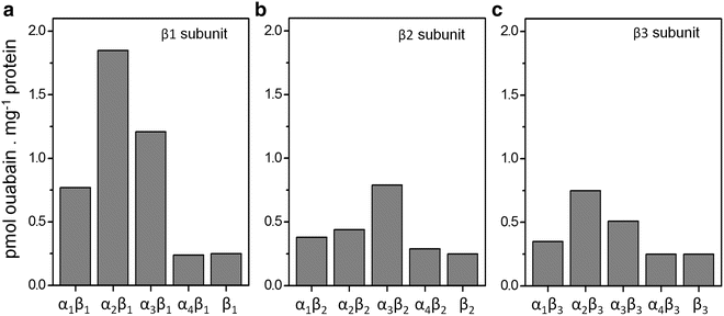
Ouabain binding of recombinant Na,K-ATPase isoforms. Na,K-ATPase α1, α2, α3, or α4 in combination with the Na,K-ATPase β1, β2, and β3 subunits are produced with the baculovirus expression system. The membrane fractions of the Sf9 cells are isolated and ouabain binding capacity of the membranes fraction is shown. Figures 2 and 3A can be compared, showing expression analyses with different methods


4 Notes
-
1.
It is important to realize that the different subunits (α and β) are produced by one virus. The use of a Gateway (Life Technologies) destination vector with constant β-subunit gives the possibility to combine it with different catalytic α-subunits. By performing mutagenesis on the pEntry-α-subunit vector the mutated α-subunit can easily be cloned into the destination vector.
-
2.
We do not determine virus titers (generally 108 pfu/mL) as this is quite laborious and experience from the past shows that directly using the volumes in this protocol gives good results.
-
3.
Viral stocks can be stored at 4 °C for many years. In our experience the titer of these stocks slowly reduces and we generally do not use stocks that are older than 2 years for protein production.
-
4.
Sf9 insect cells are cultured without CO2.
-
5.
For production of the ATPase subunits 1.0–1.5 × 10⁶ cells/mL are infected at a multiplicity of infection of 1–3 in Xpress medium.
-
6.
Many different recombinant baculoviruses expressing Na,K-ATPase [1], gastric [7] or non-gastric H+,K+-ATPase [2], or Na,K-ATPase of Drosophila melanogaster [8] have been used in the past.
References
Koenderink JB, Hermsen HP, Swarts HG, Willems PH, De Pont JJ (2000) High-affinity ouabain binding by a chimeric gastric H, K-ATPase containing transmembrane hairpins M3–M4 and M5–M6 of the alpha 1-subunit of rat Na, K-ATPase. Proc Natl Acad Sci U S A 97(21):11209–11214
Swarts HG, Koenderink JB, Willems PH, De Pont JJ (2005) The non-gastric H, K-ATPase is oligomycin-sensitive and can function as an H, NH4-ATPase. J Biol Chem 280(39):33115–33122
Luckow VA, Lee SC, Barry GF, Olins PO (1993) Efficient generation of infectious recombinant baculoviruses by site-specific transposon-mediated insertion of foreign genes into a baculovirus genome propagated in Escherichia coli. J Virol 67(8):4566–4579
Klaassen CH, Swarts HG, De Pont JJ (1995) Ethanol stimulates expression of functional H, K-ATPase in SF9 cells. Biochem Biophys Res Commun 210(3):907–913
Koenderink JB, Geibel S, Grabsch E, De Pont JJ, Bamberg E, Friedrich T (2003) Electrophysiological analysis of the mutated Na, K-ATPase cation binding pocket. J Biol Chem 278(51):51213–51222
De Pont JJ, Swarts HG, Karawajczyk A, Schaftenaar G, Willems PH, Koenderink JB (2009) The non-gastric H,K-ATPase as a tool to study the ouabain-binding site in Na,K-ATPase. Pflugers Arch 457(3):623–634
Swarts HG, Klaassen CH, de Boer M, Fransen JA, De Pont JJ (1996) Role of negatively charged residues in the fifth and sixth transmembrane domains of the catalytic subunit of gastric H,K-ATPase. J Biol Chem 271(47):29764–29772
Dalla S, Swarts HG, Koenderink JB, Dobler S (2013) Amino acid substitutions of Na,K-ATPase conferring decreased sensitivity to cardenolides in insects compared to mammals. Insect Biochem Mol Biol 43(12):1109–1115
Author information
Authors and Affiliations
Corresponding author
Editor information
Editors and Affiliations
Rights and permissions
Copyright information
© 2016 Springer Science+Business Media New York
About this protocol
Cite this protocol
Koenderink, J.B., Swarts, H.G.P. (2016). Expression of Na,K-ATPase and H,K-ATPase Isoforms with the Baculovirus Expression System. In: Bublitz, M. (eds) P-Type ATPases. Methods in Molecular Biology, vol 1377. Humana Press, New York, NY. https://doi.org/10.1007/978-1-4939-3179-8_8
Download citation
DOI: https://doi.org/10.1007/978-1-4939-3179-8_8
Publisher Name: Humana Press, New York, NY
Print ISBN: 978-1-4939-3178-1
Online ISBN: 978-1-4939-3179-8
eBook Packages: Springer Protocols