
Abstract
Arthritis affects millions of people, specifically in our aging society. Treatment can often be difficult and includes physical therapy, medicine, injections, and surgery. This chapter provides examples of administration of joint injections, showcasing anatomy, procedures, and agents used. In this brief outline, we will focus on knee, shoulder, elbow, and wrist injections.
Access provided by Autonomous University of Puebla. Download chapter PDF
Keywords
- Knee injections: Indications
- Knee injections: Agents used
- Knee injections: Midpatellar and anterior approaches
- Knee injections: Complications
- Epicondyle injection: Positioning and technique
- Golfer’s elbow: Epicondylar injection techniques and agents
- Golfer’s Elbow: Epicondylar Injection complications
- Olecranon bursitis: Causes
- Olecranon bursitis: Technique of Injection
- Olecranon bursitis: Complications of Injection
- Carpal Tunnel syndrome: General considerations—Anatomy and treatment
- Shoulder joint: Positioning and technique for injection
- Shoulder joint injections: Complications
Introduction
In this chapter, we are briefly covering some commonly used peripheral injections. It is recommended to use other resources to expand your knowledge, as there are numerous injections that may help manage your patients. Prior to proceeding with any injection full medical history, allergies, and full medication list should be overviewed prior to assure no contraindications. Proper sterile technique is recommended on all below injections.
Fluoroscopy or ultrasound can aid in precise delivery of injectate in obese or complicated patients and help to avoid sensitive structures.
Knee
Knee Intra-articular Injection
-
Indications:
-
Knee pain
-
Osteoarthritis
-
Rheumatoid arthritis
-
Gout
-
-
Injection technique:
Two different approaches: midpatellar and anterior
-
1.
Midpatellar approach: patient supine with knee extended on a pillow
-
Lateral approach: intersection of a line drawn between the lateral and proximal border of the patella (Fig. 47.1), needle inserted between the patella, and the femur at the intersection point directed at a 45° angle toward the middle of the medial side of the joint
Fig. 47.1 
Intra-articular knee injection. Lateral approach. Note that needle is inserted between the patella and the femur at a 45° angle
-
Medial approach: needle entry point from the medial side of the knee under the middle of the patella and aiming to the opposite patellar mid-pole (Fig. 47.2)
Fig. 47.3 
Intra-articular shoulder injection
-
2. Anterior approach:
-
Infrapatellar (medial or lateral approach)
-
Patient positioning: seated on the exam table allowing gravity to facilitate opening of the joint space, Knees flexed 60–90°
-
Palpate the joint space opening, mark this with a marking pen or make an impression with a retractable pen
-
Clean with betadine × 3 or chlorhexidine to prep the area of insertion
-
-
Needle is directed through insertion site either medially or laterally to the joint space opening (Fig. 47.3)
Fig. 47.4 
Injection for Carpal Tunnel syndrome. Boundaries of the Carpal Tunnel. Palmaris longus tendon and flexor retinaculum are noted here
-
Suprapatellar
Fig. 47.2 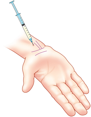
Intra-articular knee injection. Medial approach. Note the needle enters from the medial side of the knee, under the patella, aiming to the opposite patellar mid-pole
-
Patient supine
-
Best used to drain large infusions
-
Ultrasound approach: patient supine, knee flexed at 20–30°
-
High-resolution transducer parallel to the tendon quadriceps femoris, then the transducer is rotated to the axial plane.
-
Largest dimension of synovial recess should be identified and should be the target of the injection
-
Using sterile technique, a 22-gauge 3.5 in. needle is advanced into the plane of the suprapatellar recess. 2 mL of iohexol should be injected to determine intra-articular spread. Once confirmed medication should be deposited.
-
-
-
-
1.
-
Medications: Depomedrol or Kenalog 40–60 mg with 1–2 cm3 of 1 % lidocaine and 1–2 cm3 of 0.25 % bupivacaine mixture total of about 5 cm3


Shoulder [1]
Posterior Intra-articular Shoulder Injection
-
Indications:
-
Arthritis of the shoulder joint
-
Shoulder pain
-
-
Patient positioning:
-
Supine or seated
-
Arm hanging by patient’s side to allow gravity to widen the joint space
-
-
Injection technique:
-
Identify the midpoint of the acromion
-
Approximately 1 in. below this midpoint is the shoulder joint space, and the intra-articular space is more lateral
-
Prep the skin overlying the posterior shoulder/subacromial region/joint space opening in sterile fashion
-
A 1½ in 25 g needle is carefully advanced through the skin and subcutaneous tissues through to the joint.
-
If bone is encountered: pull back slightly and redirect with slight increased superior or medial tilt to proceed properly through the joint.
-
If resistance is encountered: the needle may be in ligament or tendon, it should be advanced properly into the joint space.
-
-
-
Medications: Depomedrol or Kenalog 40–60 mg with 1–2 cm3 of 1 % lidocaine and 1–2 cm3 of 0.25 % bupivacaine mixture total of about 5 cm3
-
Complications:
-
Infection: exceedingly rare with proper sterile technique
-
Transient increase in pain following injection
-
Ecchymosis and hematoma formation: decreased if pressure is placed on the injection site immediately following injection.
-
Elbow
Epicondyle Injections
Tennis Elbow (Lateral Epicondylitis) [1, 2, 4]
-
Patient positioning:
-
Patient supine or seated
-
Arm placed on exam table with affected epicondyle exposed, elbow bent
-
Palmar aspect of hand resting on the table or a folded towel to relax the affected tendons.
-
-
Injection Technique
-
Identify and mark the lateral epicondyle
-
Prep the skin in sterile fashion
-
Insert a 1 in. 25-gauge needle perpendicularly to the lateral epicondyle into the subcutaneous tissue overlying the affected tendon.
-
If bone is encountered: pull back needle slightly before injecting medications
-
If resistance is encountered: the needle may be in the tendon and should be withdrawn until the injection proceeds without significant resistance.
-
-
-
Medications: Total mixture of 2–3 mL: 1–2 mL of local anesthetic mixed with 20–40 mg of Depomedrol or Kenalog
Golfer’s Elbow (Medial Epicondylitis) [1, 6]
-
Patient positioning:
-
Patient supine or in seated position
-
Arm fully adducted with effected epicondyle exposed, elbow fully extended
-
Dorsum of the hand resting on the exam table or a folded towel to relax the affected tendons
-
-
Injection technique :
-
Identify and mark the medial epicondyle
-
Prep the skin in sterile fashion
-
Insert a 1 in. 25 g needle perpendicularly to the medial epicondyle into the subcutaneous tissue overlying the affected tendon.
-
If bone is encountered: pull back needle slightly before injecting medications
-
If resistance is encountered: the needle may be in the tendon and should be withdrawn until the injection proceeds without significant resistance.
-
-
-
Medications: Total mixture of 2–3 mL: 1–2 mL of local anesthetic mixed with 20–40 mg of Depomedrol or Kenalog
Epicondyle Injection Complications
-
Radial tunnel syndrome (lateral epicondyle injection)
-
Ulnar nerve injury at the medial ulnar groove (medial epicondyle injection)
-
Bursa injection/irritation
-
Rupture/injury of the affected tendons can result from:
-
Direct injection into the tendon which increases risk of tendinitis or tendon rupture
-
-
Infection
-
Transient increase in pain following the injection in approximately 25 % of patients
Olecranon Bursitis [7–10]
-
Activities causing this pathology
-
Direct trauma to the elbow from playing sports such as hockey or falling directly onto the olecranon process.
-
Repeated microtrauma from leaning on the elbow when arising or from working long hours at a drafting table
-
In rare cases, infection such as gout or bacterial infection
-
-
Patient positioning:
-
Supine position
-
Arm fully adducted at the patient’s side
-
Elbow flexed
-
Palm of the hand resting on the patient’s abdomen or exam table
-
-
Injection technique :
-
Identify the olecranon process and overlying bursa
-
Prep the skin overlying the posterior aspect of the joint in sterile fashion
-
Insert a 1 in. 25 g needle through the skin and subcutaneous tissues directly into the bursa
-
If bone is encountered, the needle is withdrawn into the bursa
-
-
-
Medications: Total of 5 mL: 2 mL of local anesthetic with 40 mg of methylprednisolone or equivalent steroid
-
Complications: infection, sterile conditions will minimize this potential complication.
-
Pearl: coexisting tendinitis may require additional treatment
Wrist
Carpal Tunnel Syndrome
-
The palmaris longus tendon is bound on dorsal and lateral surfaces by the carpal bones and intercarpal joints
-
The carpel tunnel is formed by the carpel bones dorsally and the transverse carpal ligament (aka flexor retinaculum) ventrally. Tendons of flexor digitorum profundus, sublimus and flexor pollicus longus, and median nerve pass through the carpal tunnel.
-
Patient positioning: Patient is supine, arm is supinated to expose inner portion of wrist
-
Injection technique: (Fig. 47.4) The injection is performed at a site just distal to the palmaris longus tendon and at the proximal wrist crease. The needle is inserted at a 30° angle and directed toward the ring finger.
-
Medications: Local corticosteroid injections into the carpal tunnel may be useful for those with CTS of short duration (less than a year). A solution of 20–40 mg of corticosteroid with 1–2 cm3 of 1 % lidocaine and 1–2 cm3 of 0.25 % bupivacaine or 0.2 % ropivacaine mixture, a total of about 5 cm3, is injected along the transverse carpal ligament.
-
Complications: Major complication: exceedingly rare if strict aseptic technique is adhered to.
Transient increase in pain following the intra-articular injection ecchymosis and hematoma formation: decreased if pressure is placed on the injection site immediately following injection.
Recommendations
-
Always follow sterile technique to avoid infection.
-
Instruct patients to avoid vigorous exercises following injection.
-
Relative rest and ice is recommended afterwards.
-
Osteoarthritis of the shoulder must be distinguished from other causes of shoulder pain including rotator cuff tear pathology to ensure proper treatment regimen.
-
Coexisting bursitis and tendinitis may contribute to shoulder pain and require additional localized injection.
-
Caution should be used when doing these injections to avoid tendon rupture/injury.
References
Waldman SD. Shoulder pain. In: Waldman SD, editor. Atlas of common pain syndromes. Philadelphia: Saunders; 2002.
Waldman S. Tennis elbow. In: Waldman S, editor. Pain management. Philadelphia: Saunders/Elsevier; 2007. p. 633–6.
Waldman SD. The tennis elbow test. In: Waldman SD, editor. Physical diagnosis of pain: an atlas of signs and symptoms. Philadelphia: Saunders; 2006.
Waldman SD. Tennis elbow. In: Waldman SD, editor. Atlas of pain management injection techniques. Philadelphia: Saunders; 2000.
Waldman SD. Golfer’s elbow. In: Waldman SD, editor. Atlas of common pain syndromes. Philadelphia: Saunders; 2002.
Waldman S. Golfer’s elbow. In: Waldman S, editor. Pain management. Philadelphia: Saunders/Elsevier; 2007. p. 637–40.
Waldman S. Olecranon cubital bursitis. In: Waldman S, editor. Pain management. Philadelphia: Saunders/Elsevier; 2007. p. 641–6.
Groff GD. Olecranon bursitis. In: Klippel JH, Dieppe PA, editors. Rheumatology. 2nd ed. London: Mosby; 1998.
McAfee JH, Smith DL. Olecranon and prepatellar bursitis: diagnosis and treatment. West J Med. 1988;149:607.
Waldman S. Olecranon cubital bursitis. In: Waldman S, editor. Atlas of common pain syndromes. Philadelphia: Saunders/Elsevier; 2007.
Author information
Authors and Affiliations
Corresponding author
Editor information
Editors and Affiliations
Rights and permissions
Copyright information
© 2015 Springer Science+Business Media New York
About this chapter
Cite this chapter
Gritsenko, K., Tomajian, S., Aquino, M., Kaye, A.D. (2015). Joint, Tendon, and Nerve Injections. In: Sackheim, K. (eds) Pain Management and Palliative Care. Springer, New York, NY. https://doi.org/10.1007/978-1-4939-2462-2_47
Download citation
DOI: https://doi.org/10.1007/978-1-4939-2462-2_47
Publisher Name: Springer, New York, NY
Print ISBN: 978-1-4939-2461-5
Online ISBN: 978-1-4939-2462-2
eBook Packages: MedicineMedicine (R0)