
Abstract
Immunohistochemistry is a powerful adjunctive technique for the pathologic diagnosis of soft tissue and bone tumors, although some tumors still lack of specific markers. This chapter includes the questions about the immunohistochemical markers for normal soft tissue and bone, soft tissue and bone tumors, and their utility for differentiation. The questions are answered in the form of tables. The photos of selected markers are also included. New markers such as claudin-5, ERG, GAP43, INI1, MDM2, MUC4, NKX2.2, NY-ESO-1, SATB2, SOX9, SOX10, STAT6, as well as other commonly used markers are discussed.
Access provided by Autonomous University of Puebla. Download chapter PDF
Keywords
- Adamantinoma
- Aggressive angiomyxoma
- Alveolar soft part sarcoma
- Angiomatoid fibrous histiocytoma
- Angiosarcoma
- Atypical fibroxanthoma
- Chordoma
- Clear cell sarcoma of soft parts
- Dermatofibrosarcoma protuberans
- Desmoplastic small round cell tumor
- Epithelioid sarcoma
- Ewing sarcoma
- Fibromatosis
- Glomangiopericytoma
- Glomus tumors
- Granular cell tumor
- Hemangioendothelioma
- Hemangiopericytoma
- Inflammatory myofibroblastic tumor
- Kaposi sarcoma
- Langerhans cell histiocytosis
- Leiomyosarcoma
- Liposarcoma
- Low-grade myofibroblastic sarcoma
- Low grade fibromyxoid sarcoma
- Malignant extrarenal rhabdoid tumor
- Malignant melanotic schwannian tumor
- Mammary-type myofibroblastoma myofibroma
- Mesenchymal chondrosarcoma
- Myoepithelial tumors
- Myxofibrosarcoma
- Neurofibroma
- Neurothekeomas
- Neuroblastoma
- Nodular fasciitis
- Notochord
- Osteosarcoma
- Ossifying fibromyxoid tumor of soft parts
- Perineurioma
- Perivascular epithelioid cell neoplasms
- Phosphaturic mesenchymal tumor
- Pleomorphic hyalinizing angiectatic tumor
- Primitive neuroectodermal tumor
- Reticulohistiocytoma
- Rhabdomyosarcoma
- Schwannoma
- Solitary fibrous tumor
- Spindle cell hemangioma
- Synovial sarcoma
- Undifferentiated pleomorphic sarcoma
- β-catenin
- Brachyury
- Calponin
- CD1a
- CD31
- CD34
- CD99
- Chromogranin A
- Claudin-1
- CDK4
- Desmin
- ERG
- FLI1
- GLUT1
- h-Caldesmon
- HHV8 LANA
- HMB-45
- INI1
- MDM2
- Melan A
- MITF
- MUC4
- myoD1
- Myogenin
- NB84
- NKX2.2
- NY-ESO-1
- S100
- SATB2
- SOX9
- SOX10
- STAT6
- TFE3
- TLE1
- Type IV collagen
- Vimentin
- WT-1
Frequently Asked Questions
-
29.1.
Summary of applications and limitations of useful markers (Table 29.1)
Table 29.1 Summary of applications and limitations of useful markers -
29.2.
Markers positive for normal soft tissue and bone (Table 29.2)
Table 29.2 Markers positive for normal soft tissue and bone
Adipocytic Tumors
-
29.3.
Markers for spindle cell lipoma/pleomorphic lipoma (Table 29.3)
Table 29.3 Markers for spindle cell lipoma/pleomorphic lipoma -
29.4.
Markers for well-differentiated liposarcoma/dedifferentiated liposarcoma (Table 29.4)
Table 29.4 Markers for well-differentiated liposarcoma/dedifferentiated liposarcoma -
29.5.
Markers for myxoid/round cell liposarcoma (Table 29.5)
Table 29.5 Markers for myxoid/round cell liposarcoma
Fibroblastic and Fibrohistiocytic Tumors
-
29.6.
Markers for nodular fasciitis (Table 29.6)
Table 29.6 Markers for nodular fasciitis -
29.7.
Markers for palmar and plantar (superficial) fibromatosis (Table 29.7)
Table 29.7 Markers for palmar and plantar (superficial) fibromatosis -
29.8.
Markers for deep fibromatosis (Table 29.8)
Table 29.8 Markers for deep fibromatosis -
29.9.
Markers for fibrous hamartoma of infancy (Table 29.9)
Table 29.9 Markers for fibrous hamartoma of infancy -
29.10.
Markers for inflammatory myofibroblastic tumor/inflammatory fibrosarcoma (Table 29.10)
Table 29.10 Markers for Inflammatory myofibroblastic tumor/inflammatory fibrosarcoma -
29.11.
Markers of myofibroma/myofibromatosis (Table 29.11)
Table 29.11 Markers of myofibroma/myofibromatosis -
29.12.
Markers for angiomyofibroblastoma (Table 29.12)
Table 29.12 Markers for angiomyofibroblastoma -
29.13.
Markers for cellular angiofibroma (Table 29.13)
Table 29.13 Markers for cellular angiofibroma -
29.14.
Markers of mammary-type myofibroblastoma (Table 29.14)
Table 29.14 Markers of mammary-type myofibroblastoma -
29.15.
Markers for myxoinflammatory fibroblastic sarcoma (Table 29.15)
Table 29.15 Markers for myxoinflammatory fibroblastic sarcoma -
29.16.
Markers for low-grade myofibroblastic sarcoma (Table 29.16)
Table 29.16 Markers for low-grade myofibroblastic sarcoma -
29.17.
Markers for low-grade fibromyxoid sarcoma including spindle cell tumor with giant rosettes (Table 29.17)
Table 29.17 Markers for low-grade fibromyxoid sarcoma/ spindle cell tumor with giant rosettes -
29.18.
Markers for myxofibrosarcoma (Table 29.18)
Table 29.18 Markers for myxofibrosarcoma
Fibrohistiocytic Tumors
-
29.19.
Markers for benign fibrous histiocytoma (Table 29.19)
Table 29.19 Markers for benign fibrous histiocytoma -
29.20.
Markers for juvenile xanthogranuloma and reticulohistiocytoma (Table 29.20)
Table 29.20 Markers for juvenile xanthogranuloma and reticulohistiocytoma -
29.21.
Markers for atypical fibroxanthoma (Table 29.21)
Table 29.21 Markers for atypical fibroxanthoma -
29.22.
Markers for dermatofibrosarcoma protuberans/giant cell fibroblastoma (Table 29.22)
Table 29.22 Markers for dermatofibrosarcoma protuberans/giant cell fibroblastoma -
29.23.
Markers for angiomatoid fibrous histiocytoma (Table 29.23)
Table 29.23 Markers for angiomatoid (malignant) fibrous histiocytoma -
29.24.
Markers for plexiform fibrohistiocytic tumor (Table 29.24)
Table 29.24 Markers for plexiform fibrohistiocytic tumor -
29.25.
Markers for giant cell tumor of soft tissue (Table 29.25)
Table 29.25 Markers for giant cell tumor of soft tissue -
29.26.
Markers for undifferentiated pleomorphic sarcoma (pleomorphic malignant fibrous histiocytoma) (Table 29.26)
Table 29.26 Markers for undifferentiated pleomorphic sarcoma (pleomorphic malignant fibrous histiocytoma)
Smooth Muscle Tumors
-
29.27.
Markers for leiomyoma and leiomyosarcoma (Table 29.27)
Table 29.27 Markers for leiomyoma and leiomyosarcoma Fig. 29.1 
(a) Leiomyosarcoma shows positive staining for desmin, (b) Leiomyosarcoma shows positive staining for h-caldesmon
-
29.28.
Markers for EBV-related leiomyosarcoma (Table 29.28)
Table 29.28 Markers for EBV-associated leiomyosarcoma

Skeletal Muscle Tumors
-
29.29.
Markers for rhabdomyoma (Table 29.29)
Table 29.29 Markers for rhabdomyoma -
29.30.
Markers for rhabdomyosarcoma (Table 29.30)
Table 29.30 Markers for rhabdomyosarcoma Fig. 29.2 
(a) Rhabdomyosarcoma shows positive staining for MyoD1, (b) Rhabdomyosarcoma shows positive staining for myogenin

Tumors of Perivascular Cells
-
29.31.
Markers for hemangiopericytoma (HPC)/solitary fibrous tumor (SFT) of soft tissue (Table 29.31)
Table 29.31 Markers for hemangiopericytoma (HPC)/solitary fibrous tumor (SFT) of soft tissue Fig. 29.3 
Solitary fibrous tumor shows positive staining for CD34
-
29.32.
Markers for myopericytoma family of tumors (infantile myofibromatosis, solitary myofibroma, infantile hemangiopericytoma, myopericytoma, glomangiopericytoma) (Table 29.32)
Table 29.32 Markers for myopericytoma family of tumors (infantile myofibromatosis, solitary myofibroma, infantile hemangiopericytoma myopericytoma, glomangiopericytoma) -
29.33.
Markers for glomus tumor (Table 29.33)
Table 29.33 Markers for glomus tumor Fig. 29.4 
(a) Glomus tumor on H&E section, (b) Glomus tumor shows positive staining for SMA


Vascular Tumors
-
29.34.
Markers for kaposiform hemangioendothelioma (Table 29.34)
Table 29.34 Markers for kaposiform hemangioendothelioma -
29.35.
Markers for hemangioma (Table 29.35)
Table 29.35 Markers for hemangioma -
29.36.
Markers for Dabska-type and retiform hemangioendothelioma (Table 29.36)
Table 29.36 Markers for Dabska-type and retiform hemangioendothelioma -
29.37.
Markers for epithelioid hemangioendothelioma (Table 29.37)
Table 29.37 Markers for epithelioid hemangioendothelioma -
29.38.
Markers for angiosarcoma (Table 29.38)
Table 29.38 Markers for angiosarcoma Fig. 29.5 
(a) Angiosarcoma shows positive staining for ERG, (b) Angiosarcoma shows positive staining for CD31
-
29.39.
Markers for Kaposi sarcoma (Table 29.39)
Table 29.39 Markers for Kaposi sarcoma -
29.40.
Markers for lymphatic tumors (Table 29.40)
Table 29.40 Markers for lymphatic tumors

Nerve Sheath and Neuroectodermal Tumors
-
29.41.
Markers for neurofibroma (Table 29.41)
Table 29.41 Markers for neurofibroma -
29.42.
Markers for schwannoma/nerve sheath myxoma (Table 29.42)
Table 29.42 Markers for schwannoma/nerve sheath myxoma Fig. 29.6 
Schwannoma shows positive staining for S100
-
29.43.
Markers for psammomatous melanotic schwannoma/malignant melanotic schwannian tumor (Table 29.43)
Table 29.43 Markers for psammomatous melanotic schwannoma/melanotic schwannian tumor -
29.44.
Markers for malignant peripheral nerve sheath tumor (MPNST) (Table 29.44)
Table 29.44 Markers for malignant peripheral nerve sheath tumor (MPNST) -
29.45.
Markers for granular cell tumor (Table 29.45)
Table 29.45 Markers for granular cell tumor -
29.46.
Markers for neurothekeomas (Table 29.46)
Table 29.46 Markers for cellular neurothekeomas (fibrohistiocytic tumor) -
29.47.
Markers for perineurioma (Table 29.47)
Table 29.47 Markers for perineurioma -
29.48.
Markers for neuroblastoma (Table 29.48)
Table 29.48 Markers for neuroblastoma Fig. 29.7 
(a) Neuroblastoma shows positive staining for CD56, (b) Neuroblastoma shows positive staining for synaptophysin
-
29.49.
Markers for clear cell sarcoma of soft parts (Table 29.49)
Table 29.49 Markers for clear cell sarcoma of soft parts


Tumors of Uncertain Differentiation
-
29.50.
Markers for myxoma (Table 29.50)
Table 29.50 Markers for myxoma -
29.51.
Markers for aggressive angiomyxoma (Table 29.51)
Table 29.51 Markers for aggressive angiomyxoma -
29.52.
Markers for ossifying fibromyxoid tumor of soft parts (Table 29.52)
Table 29.52 Markers for ossifying fibromyxoid tumor of soft parts -
29.53.
Markers for myoepithelial tumors (mixed tumor/myoepithelioma/parachordoma) (Table 29.53)
Table 29.53 Markers for mixed tumor/myoepithelioma/parachordoma(myoepithelial tumors) -
29.54.
Markers for pleomorphic hyalinizing angiectatic tumor (Table 29.54)
Table 29.54 Markers for pleomorphic hyalinizing angiectatic tumor -
29.55.
Markers for phosphaturic mesenchymal tumor (Table 29.55)
Table 29.55 Markers for phosphaturic mesenchymal tumor -
29.56.
Markers for perivascular epithelioid cell neoplasms (angiomyolipoma of the kidney or other organs, clear cell sugar tumor of the lung, lymphangioleiomyomatosis, clear cell myomelanocytic tumor of the falciform ligament/ligamentum teres) (Table 29.56)
Table 29.56 Markers for perivascular epithelioid cell family of tumors (PEComas) (angiomyolipoma of the kidney or other organs, clear cell sugar tumor of the lung, lymphangioleiomyomatosis, clear cell myomelanocytic tumor of the falciform ligament/ligamentum teres) -
29.57.
Markers for epithelioid sarcoma (Table 29.57)
Table 29.57 Markers for epithelioid sarcoma Fig. 29.8 
(a) Epithelioid sarcoma on H&E section, (b) Epithelioid sarcoma shows no nuclear staining for INI1, (c) Epithelioid sarcoma shows positive staining for AE1/AE3, (d) Epithelioid sarcoma shows positive staining for EMA
-
29.58.
Markers for alveolar soft part sarcoma (Table 29.58)
Table 29.58 Markers for alveolar soft part sarcoma -
29.59.
Markers for Ewing sarcoma and primitive neuroectodermal tumor (ES/PNET) (Table 29.59)
Table 29.59 Markers for Ewing sarcoma and primitive neuroectodermal tumor (ES/PNET) Fig. 29.9 
(a) Ewing sarcoma shows positive staining for FLI1, (b) Ewing sarcoma/PNENT shows positive staining for NKX2.2
-
29.60.
Markers for synovial sarcoma (Table 29.60)
Table 29.60 Markers for synovial sarcoma Fig. 29.10 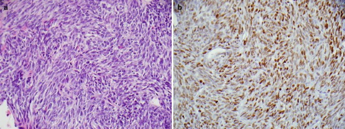
(a) Synovial sarcoma on H&E section, (b) Synovial sarcoma shows positive staining for TLE1
-
29.61.
Markers for desmoplastic small round cell tumor (Table 29.61)
Table 29.61 Markers for desmoplastic small round cell tumor Fig. 29.11 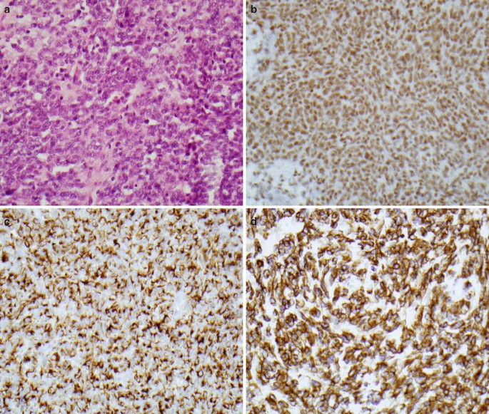
(a) DSRCT on H&E section, (b) DSRCT shows positive staining for WT1, (c) DSRCT shows positive staining for desmin, (d) DSRCT shows positive staining for AE1/AE3
-
29.62.
Markers for malignant extrarenal rhabdoid tumor (Table 29.62)
Table 29.62 Markers for malignant extrarenal rhabdoid tumor


Bone Tumors
-
29.63.
Markers for mesenchymal chondrosarcoma (Table 29.63)
Table 29.63 Markers for mesenchymal chondrosarcoma -
29.64.
Markers for chordoma (Table 29.64)
Table 29.64 Markers for chordoma -
29.65.
Markers for adamantinoma (Table 29.65)
Table 29.65 Markers for adamantinoma -
29.66.
Markers for Langerhans cell histiocytosis (Table 29.66)
Table 29.66 Markers for Langerhans cell histiocytosis
Differential Diagnosis
-
29.67.
Inflammatory myofibroblastic tumor (IMT)/inflammatory fibrosarcoma (IF) vs. dendritic cell neoplasms (DCN) (Table 29.67)
Table 29.67 Inflammatory myofibroblastic tumor (IMT)/inflammatory fibrosarcoma (IF) vs. dendritic reticulum cell tumor (DCT) -
29.68.
Inflammatory myofibroblastic tumor (IMT)/inflammatory fibrosarcoma (IF) vs. sarcomatoid urothelial carcinoma (SUC) (Table 29.68)
Table 29.68 Inflammatory myofibroblastic tumor (IMT)/inflammatory fibrosarcoma (IF) vs. sarcomatoid urothelial carcinoma (SUC) -
29.69.
Myxoinflammatory fibroblastic sarcoma (MFS) vs. Hodgkin’s lymphoma (HL) (Table 29.69)
Table 29.69 Myxoinflammatory fibroblastic sarcoma (MFS) vs. Hodgkin’s lymphoma (HL) -
29.70.
Fibrosarcoma (CF) vs. monomorphic synovial sarcoma (MSS) vs. malignant peripheral nerve sheath tumor (MPNST) vs. spindle cell carcinoma (SCC) vs. spindle cell melanoma (SCM) vs. dermatofibrosarcoma protuberans (DFSP) vs. spindle cell rhabdomyosarcoma (SCR) vs. leiomyosarcoma (LMS) vs. spindle cell angiosarcoma (SCA) (Table 29.70)
Table 29.70 Fibrosarcoma (CF) vs. monomorphic synovial sarcoma (MSS) vs. malignant peripheral nerve sheath tumor (MPNST) vs. spindle cell carcinoma (SCC) vs. spindle cell melanoma (SCM) vs. Dermatofibrosarcoma protuberans (DFSP) vs. spindle cell rhabdomyosarcoma (SCR) vs. leiomyosarcoma (LMS) vs. spindle cell angiosarcoma (SCA) -
29.71.
Low grade fibromyxoid sarcoma (LGFS) vs. neurofibroma/perineurioma vs. desmoid tumors (Table 29.71)
Table 29.71 Low grade fibromyxoid sarcoma (LGFS) vs. neurofibroma/perineurioma vs. desmoid tumors -
29.72.
Juvenile xanthogranuloma (JXG)/reticulohistiocytoma (RC) vs. Langerhans cell histiocytosis (LCH) (Table 29.72)
Table 29.72 Juvenile xanthogranuloma (JXG)/reticulohistiocytoma (RC) vs. Langerhans cell histiocytosis (LCH) -
29.73.
Dermatofibroma protuberans (DFSP) vs. dermatofibroma (DF) vs. fibrosarcoma arising in DFSP (Table 29.73)
Table 29.73 Dermatofibroma protuberans (DFSP) vs. dermatofibroma (DF) vs. fibrosarcoma arising in DFSP -
29.74.
Rhabdomyosarcoma (RMS) vs. Ewing sarcoma/PNET (ES/PNET) vs. neuroblastoma (NB) vs. desmoplastic small round cell tumor (DSRCT) vs. small cell synovial sarcoma (SCSS) vs. lymphoma (Table 29.74)
Table 29.74 Rhabdomyosarcoma (RMS) vs. Ewing sarcoma/PNET (ES/PNET) vs. neuroblastoma (NB) vs. desmoplastic small round cell tumor (DSRCT) vs. synovial sarcoma (SS) vs. lymphoma -
29.75.
Leiomyoma vs. GIST vs. schwannoma (Table 29.75)
Table 29.75 Leiomyoma vs. GIST vs. schwannoma -
29.76.
Glomus tumors (GT) vs. hemangiopericytoma (HPC)/solitary fibrous tumor (SFT) of soft tissue (Table 29.76)
Table 29.76 Glomus tumors (GT) vs. hemangiopericytoma (HPC)/solitary fibrous tumor (SFT) of soft tissue -
29.77.
Kaposiform hemangioendothelioma (KH) vs. capillary hemangioma (CH) (Table 29.77)
Table 29.77 Kaposiform hemangioendothelioma (KH) vs. juvenile capillary hemangioma (CH) -
29.78.
Neurofibroma vs. schwannoma vs. perineurioma (Table 29.78)
Table 29.78 Neurofibroma vs. schwannoma vs. perineurioma -
29.79.
ES/PNET vs. lymphoblastic lymphoma (Table 29.79)
Table 29.79 ES/PNET vs. lymphoblastic lymphoma (LL) -
29.80.
ES/PNET vs. neuroblastoma (Table 29.80)
Table 29.80 ES/PNET vs. neuroblastoma -
29.81.
Chordoma vs. chondrosarcoma (Table 29.81)
Table 29.81 Chordoma vs. chondrosarcoma -
29.82.
Chordoma vs. renal cell carcinoma (Table 29.82)
Table 29.82 Chordoma vs. renal cell carcinoma -
29.83.
Langerhans cell histiocytosis (LCH) vs. Hodgkin lymphoma (HL) (Table 29.83)
Table 29.83 Langerhans’ cell histiocytosis (LCH) vs. Hodgkin lymphoma (HL) -
29.84.
Epithelioid sarcoma (ES) vs. epithelioid angiosarcoma (EA) vs. epithelioid malignant peripheral nerve sheath tumor (EPNST) vs. epithelioid leiomyosarcoma (ELMS) vs. sclerosing epithelioid fibrosarcoma (SEF) vs. epithelioid osteosarcoma (EO) (Table 29.84)
Table 29.84 Epithelioid sarcoma (ES) vs. epithelioid angiosarcoma (EA) vs. epithelioid malignant peripheral nerve sheath tumor (EPNST) vs. epithelioid leiomyosarcoma (ELMS) vs. sclerosing epithelioid fibrosarcoma (SEF) vs. epithelioid osteosarcoma (EO) -
29.85.
Rhabdomyoma vs. granular cell tumor (Table 29.85)
Table 29.85 Rhabdomyoma vs. granular cell tumor -
29.86.
Rhabdomyosarcoma vs. leiomyosarcoma (Table 29.86)
Table 29.86 Rhabdomyosarcoma vs. leiomyosarcoma
Adipocytic Tumors
Fibroblastic and Fibrohistiocytic Tumors
Fibrohistiocytic tumors
Smooth Muscle Tumors
Tumors of Perivascular Cells
References
Dabbs DJ. Diagnostic immunohistochemistry. 4th edn. Philadelphia, PA: Churchill Livingstone Elsevier; 2014. Accessed 1 May 2014.
Fletcher CD, Unni KK, Mertens F. WHO classification of tumours: pathology and genetics: tumours of soft tissue and bone. Lyon: IARC Press; 2002. 427.
Folpe AL, Inwards CY. Foundations in diagnostic pathology: bone and soft tissue pathology. Philadelphia, PA: Saunders Elsevier; 2010. p. 462.
Weiss SW, Goldblum JR. Enzinger and Weiss’s soft tissue tumors. 5th edn. Philadelphia, PA: Mosby Elsevier; 2008, p 1257. Accessed 21 May 2010.
Suster S, Fisher C. Immunoreactivity for the human hematopoietic progenitor cell antigen (CD34) in lipomatous tumors. Am J Surg Pathol. 1997;21(2):195–200.
Templeton SF, Solomon Jr AR. Spindle cell lipoma is strongly CD34 positive. An immunohistochemical study. J Cutan Pathol. 1996;23(6):546–50.
Suster S, Fisher C, Moran CA. Expression of bcl-2 oncoprotein in benign and malignant spindle cell tumors of soft tissue, skin, serosal surfaces, and gastrointestinal tract. Am J Surg Pathol. 1998;22(7):863–72.
Thway K, Flora R, Shah C. Diagnostic utility of p16, CDK4, and MDM2 as an immunohistochemical panel in distinguishing well-differentiated and dedifferentiated liposarcomas from other adipocytic tumors. Am J Surg Pathol. 2012;36(3):462–9.
Haimoto H, Kato K, Suzuki F, Nagura H. The ultrastructural changes of S-100 protein localization during lipolysis in adipocytes. An immunoelectron-microscopic study. Am J Pathol. 1985;121(2):185–91.
Binh MB, Garau XS, Guillou L, Aurias A, Coindre JM. Reproducibility of MDM2 and CDK4 staining in soft tissue tumors. Am J Clin Pathol. 2006;125(5):693–7.
Binh MB, Sastre-Garau X, Guillou L, et al. MDM2 and CDK4 immunostainings are useful adjuncts in diagnosing well-differentiated and dedifferentiated liposarcoma subtypes: a comparative analysis of 559 soft tissue neoplasms with genetic data. Am J Surg Pathol. 2005;29(10):1340–7.
Hostein I, Pelmus M, Aurias A, Pedeutour F, Mathoulin-Pelissier S, Coindre JM. Evaluation of MDM2 and CDK4 amplification by real-time PCR on paraffin wax-embedded material: a potential tool for the diagnosis of atypical lipomatous tumours/well-differentiated liposarcomas. J Pathol. 2004;202(1):95–102.
Mariño-Enríquez A, Fletcher CD, Dal Cin P, et al. Dedifferentiated Liposarcoma with “homologous” lipoblastic (pleomorphic liposarcoma-like) differentiation: clinicopathologic and molecular analysis of a series suggesting revised diagnostic criteria. Am J Surg Pathol. 2010;34:1122–31.
De Vreeze RS, de Jong D, Tielen IH, et al. Primary retroperitoneal myxoid/round cell liposarcoma is a nonexisting disease: an immunohistochemical and molecular biological analysis. Mod Pathol. 2008;22:223–31.
Hisaoka M, Tsuji S, Morimitsu Y, et al. Detection of TLS/FUS-CHOP fusion transcripts in myxoid and round cell liposarcomas by nested reverse transcription-polymerase chain reaction using archival paraffin-embedded tissues. Diagn Mol Pathol. 1998;7(2):96–101.
Oikawa K, Ishida T, Imamura T, et al. Generation of the novel monoclonal antibody against TLS/EWS-CHOP chimeric oncoproteins that is applicable to one of the most sensitive assays for myxoid and round cell liposarcomas. Am J Surg Pathol. 2006;30(3):351–6.
Hemminger JA, Iwenofu OH. NY-ESO-1 is a sensitive and specific immunohistochemical marker for myxoid and round cell liposarcomas among related mesenchymal myxoid neoplasms. Mod Pathol. 2013;26:1204–10.
Montgomery EA, Meis JM. Nodular fasciitis. Its morphologic spectrum and immunohistochemical profile. Am J Surg Pathol. 1991;15(10):942–8.
Bhattacharya B, Dilworth HP, Iacobuzio-Donahue C, et al. Nuclear beta-catenin expression distinguishes deep fibromatosis from other benign and malignant fibroblastic and myofibroblastic lesions. Am J Surg Pathol. 2005;29(5):653–9.
Carlson JW, Fletcher CD. Immunohistochemistry for beta-catenin in the differential diagnosis of spindle cell lesions: analysis of a series and review of the literature. Histopathology. 2007;51(4):509–14.
Saab ST, McClain CM, Coffin CM. Fibrous hamartoma of infancy a clinicopathologic analysis of 60 cases. Am J Surg Pathol. 2014;38:394–401.
Ceballos KM, Nielsen GP, Selig MK, O’Connell JX. Is anti-h-caldesmon useful for distinguishing smooth muscle and myofibroblastic tumors? an immunohistochemical study. Am J Clin Pathol. 2000;114(5):746–53.
Coffin CM, Hornick JL, Fletcher CD. Inflammatory myofibroblastic tumor: comparison of clinicopathologic, histologic, and immunohistochemical features including ALK expression in atypical and aggressive cases. Am J Surg Pathol. 2007;31(4):509–20.
Coffin CM, Watterson J, Priest JR, Dehner LP. Extrapulmonary inflammatory myofibroblastic tumor (inflammatory pseudotumor). A clinicopathologic and immunohistochemical study of 84 cases. Am J Surg Pathol. 1995;19(8):859–72.
Cook JR, Dehner LP, Collins MH, et al. Anaplastic lymphoma kinase (ALK) expression in the inflammatory myofibroblastic tumor: a comparative immunohistochemical study. Am J Surg Pathol. 2001;25(11):1364–71.
Harik LR, Merino C, Coindre JM, Amin MB, Pedeutour F, Weiss SW. Pseudosarcomatous myofibroblastic proliferations of the bladder: a clinicopathologic study of 42 cases. Am J Surg Pathol. 2006;30(7):787–94.
Daimaru Y, Hashimoto H, Enjoji M. Myofibromatosis in adults (adult counterpart of infantile myofibromatosis). Am J Surg Pathol. 1989;13(10):859–65.
Fletcher CD, Tsang WY, Fisher C, Lee KC, Chan JK. Angiomyofibroblastoma of the vulva. A benign neoplasm distinct from aggressive angiomyxoma. Am J Surg Pathol. 1992;16(4):373–82.
Laskin WB, Fetsch JF, Mostofi FK. Angiomyofibroblastomalike tumor of the male genital tract: analysis of 11 cases with comparison to female angiomyofibroblastoma and spindle cell lipoma. Am J Surg Pathol. 1998;22(1):6–16.
Laskin WB, Fetsch JF, Tavassoli FA. Angiomyofibroblastoma of the female genital tract: analysis of 17 cases including a lipomatous variant. Hum Pathol. 1997;28(9):1046–55.
Nielsen GP, Rosenberg AE, Young RH, Dickersin GR, Clement PB, Scully RE. Angiomyofibroblastoma of the vulva and vagina. Mod Pathol. 1996;9(3):284–91.
Ockner DM, Sayadi H, Swanson PE, Ritter JH, Wick MR. Genital angiomyofibroblastoma. Comparison with aggressive angiomyxoma and other myxoid neoplasms of skin and soft tissue. Am J Clin Pathol. 1997;107(1):36–44.
Iwasa Y, Fletcher CD. Cellular angiofibroma: clinicopathologic and immunohistochemical analysis of 51 cases. Am J Surg Pathol. 2004;28(11):1426–35.
Lee AH, Sworn MJ, Theaker JM, Fletcher CD. Myofibroblastoma of breast: an immunohistochemical study. Histopathology. 1993;22(1):75–8.
Meis-Kindblom JM, Kindblom LG. Acral myxoinflammatory fibroblastic sarcoma: a low-grade tumor of the hands and feet. Am J Surg Pathol. 1998;22(8):911–24.
Laskin WB, Fetsch JF, Miettinen M. Myxoinflammatory fibroblastic sarcoma: a clinicopathologic analysis of 104 cases, with emphasis on predictors of outcome. Am J Surg Pathol. 2014;38(1):1–12.
Mentzel T, Dry S, Katenkamp D, Fletcher CD. Low-grade myofibroblastic sarcoma: analysis of 18 cases in the spectrum of myofibroblastic tumors. Am J Surg Pathol. 1998;22(10):1228–38.
Goodlad JR, Mentzel T, Fletcher CD. Low grade fibromyxoid sarcoma: clinicopathological analysis of eleven new cases in support of a distinct entity. Histopathology. 1995;26(3):229–37.
Lane KL, Shannon RJ, Weiss SW. Hyalinizing spindle cell tumor with giant rosettes: a distinctive tumor closely resembling low-grade fibromyxoid sarcoma. Am J Surg Pathol. 1997;21(12):1481–8.
Doyle LA, Moller E, Cin DP, et al. MUC4 is a highly sensitive and specific marker for low-grade fibromyxoid sarcoma. Am J Surg Pathol. 2011;35:733–41.
Mentzel T, Calonje E, Wadden C, et al. Myxofibrosarcoma. Clinicopathologic analysis of 75 cases with emphasis on the low-grade variant. Am J Surg Pathol. 1996;20(4):391–405.
Fukunaga M, Fukunaga N. Low-grade myxofibrosarcoma: progression in recurrence. Pathol Int. 1997;47(2–3):161–5.
Smith SC, Poznanski AA, Fullen DR. CD34-positive superficial myxofibrosarcoma: a potential diagnostic pitfall. J Cutan Pathol. 2013;40(7):639–45.
Nascimento AG. A clinicopathologic and immunohistochemical comparative study of cutaneous and intramuscular forms of juvenile xanthogranuloma. Am J Surg Pathol. 1997;21(6):645–52.
Sonoda T, Hashimoto H, Enjoji M. Juvenile xanthogranuloma. Clinicopathologic analysis and immunohistochemical study of 57 patients. Cancer. 1985;56(9):2280–6.
Kanner WA, Brill II LB, Patterson JW, Wick MR. CD10, p63 and CD99 expression in the differential diagnosis of atypical fibroxanthoma, spindle cell squamous cell carcinoma and desmoplastic melanoma. J Cutan Pathol. 2010;37:744–50.
Longacre TA, Smoller BR, Rouse RV. Atypical fibroxanthoma. Multiple immunohistologic profiles. Am J Surg Pathol. 1993;17(12):1199–209.
Altman DA, Nickoloff BJ, Fivenson DP. Differential expression of factor XIIIa and CD34 in cutaneous mesenchymal tumors. J Cutan Pathol. 1993;20(2):154–8.
Goldblum JR, Reith JD, Weiss SW. Sarcomas arising in dermatofibrosarcoma protuberans: a reappraisal of biologic behavior in eighteen cases treated by wide local excision with extended clinical follow up. Am J Surg Pathol. 2000;24(8):1125–30.
Fletcher CD. Angiomatoid “malignant fibrous histiocytoma”: an immunohistochemical study indicative of myoid differentiation. Hum Pathol. 1991;22(6):563–8.
Smith ME, Costa MJ, Weiss SW. Evaluation of CD68 and other histiocytic antigens in angiomatoid malignant fibrous histiocytoma. Am J Surg Pathol. 1991;15(8):757–63.
Fanburg-Smith JC, Miettinen M. Angiomatoid “malignant” fibrous histiocytoma: a clinicopathologic study of 158 cases and further exploration of the myoid phenotype. Hum Pathol. 1999;30(11):1336–43.
Schaefer IM, Fletcher CD. Myxoid variant of so-called angiomatoid “malignant fibrous histiocytoma”: clinicopathologic characterization in a series of 21 cases. Am J Surg Pathol. 2014;38:816–23.
Hollowood K, Holley MP, Fletcher CD. Plexiform fibrohistiocytic tumour: clinicopathological, immunohistochemical and ultrastructural analysis in favor of a myofibroblastic lesion. Histopathology. 1991;19(6):503–13.
Moosavi C, Jha P, Fanburg-Smith JC. An update on plexiform fibrohistiocytic tumor and addition of 66 new cases from the Armed Forces Institute of Pathology, in honor of Franz M. Enzinger, MD. Ann Diagn Pathol. 2007;11(5):313–9.
Remstein ED, Arndt CA, Nascimento AG. Plexiform fibrohistiocytic tumor: clinicopathologic analysis of 22 cases. Am J Surg Pathol. 1999;23(6):662–70.
O’Connell JX, Wehrli BM, Nielsen GP, Rosenberg AE. Giant cell tumors of soft tissue: a clinicopathologic study of 18 benign and malignant tumors. Am J Surg Pathol. 2000;24(3):386–95.
Coindre JM, Hostein I, Maire G, et al. Inflammatory malignant fibrous histiocytomas and dedifferentiated liposarcomas: histological review, genomic profile, and MDM2 and CDK4 status favour a single entity. J Pathol. 2004;203(3):822–30.
Fletcher CD, Gustafson P, Rydholm A, Willen H, Akerman M. Clinicopathologic re-evaluation of 100 malignant fibrous histiocytomas: prognostic relevance of subclassification. J Clin Oncol. 2001;19(12):3045–50.
Khalidi HS, Singleton TP, Weiss SW. Inflammatory malignant fibrous histiocytoma: distinction from Hodgkin’s disease and non-Hodgkin’s lymphoma by a panel of leukocyte markers. Mod Pathol. 1997;10(5):438–42.
Nielsen GP, Rosenberg AE, Koerner FC, Young RH, Scully RE. Smooth-muscle tumors of the vulva. A clinicopathological study of 25 cases and review of the literature. Am J Surg Pathol. 1996;20(7):779–93.
Paal E, Miettinen M. Retroperitoneal leiomyomas: a clinicopathologic and immunohistochemical study of 56 cases with a comparison to retroperitoneal leiomyosarcomas. Am J Surg Pathol. 2001;25(11):1355–63.
Perez-Montiel MD, Plaza JA, Dominguez-Malagon H, Suster S. Differential expression of smooth muscle myosin, smooth muscle actin, h-caldesmon, and calponin in the diagnosis of myofibroblastic and smooth muscle lesions of skin and soft tissue. Am J Dermatopathol. 2006;28(2):105–11.
Jenson HB, Montalvo EA, McClain KL, et al. Characterization of natural Epstein-Barr virus infection and replication in smooth muscle cells from a leiomyosarcoma. J Med Virol. 1999;57(1):36–46.
Kapadia SB, Meis JM, Frisman DM, Ellis GL, Heffner DK. Fetal rhabdomyoma of the head and neck: a clinicopathologic and immunophenotypic study of 24 cases. Hum Pathol. 1993;24(7):754–65.
Martin SE, Temm CJ, Goheen PM, et al. Cytoplasmic p63 immunohistochemistry is a useful marker for muscle differentiation: an immunohistochemical and immunoelectron microscopic study. Mod Pathol. 2011;24:1320–6.
Miettinen M, Rapola J. Immunohistochemical spectrum of rhabdomyosarcoma and rhabdomyosarcoma-like tumors. Am J Surg Pathol. 1989;13(2):120–32.
Folpe AL. MyoD1 and myogenin expression in human neoplasia: a review and update. Adv Anat Pathol. 2002;9(3):198–203.
Parham DM, Webber B, Holt H, Williams WK, Maurer H. Immunohistochemical study of childhood rhabdomyosarcomas and related neoplasms. Results of an Intergroup Rhabdomyosarcoma study project. Cancer. 1991;67(12):3072–80.
Wexler LH, Ladanyi M. Diagnosing alveolar rhabdomyosarcoma: morphology must be coupled with fusion confirmation. J Clin Oncol. 2010;28(13):2126–8.
Williamson D, Missiaglia E, de Reynies A, et al. Fusion gene-negative alveolar rhabdomyosarcoma is clinically and molecularly indistinguishable from embryonal rhabdomyosarcoma. J Clin Oncol. 2010;28(13):2151–8.
Renshaw AA. O13 (CD99) in spindle cell tumors. Reactivity with hemangiopericytoma, solitary fibrous tumor, synovial sarcoma, and meningioma but rarely with sarcomatoid mesothelioma. Appl Immunohistochem. 1995;3:250–6.
Tihan T, Viglione M, Rosenblum MK, Olivi A, Burger PC. Solitary fibrous tumors in the central nervous system. A clinicopathologic review of 18 cases and comparison to meningeal hemangiopericytomas. Arch Pathol Lab Med. 2003;127(4):432–9.
van de Rijn M, Lombard CM, Rouse RV. Expression of CD34 by solitary fibrous tumors of the pleura, mediastinum, and lung. Am J Surg Pathol. 1994;18(8):814–20.
Shidham VB, Chivukula M, Gupta D, Rao RN, Komorowski R. Immunohistochemical comparison of gastrointestinal stromal tumor and solitary fibrous tumor. Arch Pathol Lab Med. 2002;126(10):1189–92.
Chilosi M, Facchettti F, Dei Tos AP, et al. Bcl-2 expression in pleural and extrapleural solitary fibrous tumours. J Pathol. 1997;181:362–7.
Middleton LP, Duray PH, Merino MJ. The histological spectrum of hemangiopericytoma: application of immunohistochemical analysis including proliferative markers to facilitate diagnosis and predict prognosis. Hum Pathol. 1998;29(6):636–40.
Rao N, Colby TV, Falconieri G, et al. Intrapulmonary solitary fibrous tumors: clinicopathologic and immunohistochemical study of 24 cases. Am J Surg Pathol. 2013;37:155–66.
Doyle LA, VIvero M, Fletcher CD, et al. Nuclear expression of STAT6 distinguishes solitary fibrous tumor from histologic mimics. Mod Pathol. 2014;27:390–5.
Matsuyama A, Hisaoka M, Hashimoto H. Angioleiomyoma: a clinicopathologic and immunohistochemical reappraisal with special reference to the correlation with myopericytoma. Hum Pathol. 2007;38(4):645–51.
Mentzel T, Dei Tos AP, Sapi Z, et al. Myopericytoma of skin and soft tissues: clinicopathologic and immunohistochemical study of 54 cases. Am J Surg Pathol. 2006;30(1):104–13.
Nuovo M, Grimes M, Knowles D. Glomus tumors: a clinicopathologic and immunohistochemical analysis of forty cases. Surg Pathol. 1990;3:31–45.
Fletcher CD, Beham A, Schmid C. Spindle cell haemangioendothelioma: a clinicopathological and immunohistochemical study indicative of a non-neoplastic lesion. Histopathology. 1991;18(4):291–301.
Folpe AL, Veikkola T, Valtola R, Weiss SW. Vascular endothelial growth factor receptor-3 (VEGFR-3): a marker of vascular tumors with presumed lymphatic differentiation, including Kaposi’s sarcoma, kaposiform and Dabska-type hemangioendotheliomas, and a subset of angiosarcomas. Mod Pathol. 2000;13(2):180–5.
Folpe AL, Chand EM, Goldblum JR, Weiss SW. Expression of Fli-1, a nuclear transcription factor, distinguishes vascular neoplasms from potential mimics. Am J Surg Pathol. 2001;25(8):1061–6.
Mentzel T, Mazzoleni G, Dei Tos AP, Fletcher CD. Kaposiform hemangioendothelioma in adults. Clinicopathologic and immunohistochemical analysis of three cases. Am J Clin Pathol. 1997;108(4):450–5.
Drut RM, Drut R. Extracutaneous infantile haemangioma is also Glut1 positive. J Clin Pathol. 2004;57(11):1197–200.
North PE, Waner M, Mizeracki A, et al. GLUT1: a newly discovered immunohistochemical marker for juvenile hemangiomas. Hum Pathol. 2000;31(1):11–22.
Calonje E, Fletcher CD, Wilson-Jones E, Rosai J. Retiform hemangioendothelioma. A distinctive form of low-grade angiosarcoma delineated in a series of 15 cases. Am J Surg Pathol. 1994;18(2):115–25.
Dabska M. Malignant endovascular papillary angioendothelioma of the skin in childhood. Clinicopathologic study of 6 cases. Cancer. 1969;24(3):503–10.
Duke D, Dvorak A, Harris TJ, Cohen LM. Multiple retiform hemangioendotheliomas. A low-grade angiosarcoma. Am J Dermatopathol. 1996;18(6):606–10.
Fanburg-Smith JC, Michal M, Partanen TA, Alitalo K, Miettinen M. Papillary intralymphatic angioendothelioma (PILA): a report of twelve cases of a distinctive vascular tumor with phenotypic features of lymphatic vessels. Am J Surg Pathol. 1999;23(9):1004–10.
Mentzel T, Beham A, Calonje E, Katenkamp D, Fletcher CD. Epithelioid hemangioendothelioma of skin and soft tissues: clinicopathologic and immunohistochemical study of 30 cases. Am J Surg Pathol. 1997;21(4):363–74.
Fletcher CD, Beham A, Bekir S, Clarke AM, Marley NJ. Epithelioid angiosarcoma of deep soft tissue: a distinctive tumor readily mistaken for an epithelial neoplasm. Am J Surg Pathol. 1991;15(10):915–24.
Miettinen M, Wang ZF, Paetau A, et al. ERG transcription factor as an immunohistochemical marker for vascular endothelial tumors and prostatic carcinoma. Am J Surg Pathol. 2011;35:432–41.
Miettinen M, Sarlomo-Rikala M, Wang ZF. Claudin-5 as an immunohistochemical marker for angiosarcoma and hemangioendotheliomas. Am J Surg Pathol. 2011;35:1848–56.
Yaskiv O, Rubin BP, He H, et al. ERG protein expression in human tumors detected with a rabbit monoclonal antibody. Am J Clin Pathol. 2012;138:803–10.
Hammock L, Reisenauer A, Wang W, Cohen C, Birdsong G, Folpe AL. Latency-associated nuclear antigen expression and human herpesvirus-8 polymerase chain reaction in the evaluation of Kaposi sarcoma and other vascular tumors in HIV-positive patients. Mod Pathol. 2005;18(4):463–8.
Guillou L, Fletcher CD. Benign lymphangioendothelioma (acquired progressive lymphangioma): a lesion not to be confused with well-differentiated angiosarcoma and patch stage Kaposi's sarcoma: clinicopathologic analysis of a series. Am J Surg Pathol. 2000;24(8):1047–57.
Kahn HJ, Bailey D, Marks A. Monoclonal antibody D2-40, a new marker of lymphatic endothelium, reacts with Kaposi’s sarcoma and a subset of angiosarcomas. Mod Pathol. 2002;15:434–40.
Galambos C, Nodit L. Identification of lymphatic endothelium in pediatric vascular tumors and malformations. Pediatr Dev Pathol. 2005;8(2):181–9.
Fukunaga M. Expression of D2-40 in lymphatic endothelium of normal tissues and in vascular tumours. Histopathology. 2005;46:396.
Miettinen M, Wang ZF. Prox1 transcription factor as a marker for vascular tumors—evaluation of 314 vascular endothelial and 1086 nonvascular tumors. Am J Surg Pathol. 2012;36:351–9.
Chen WS, Chen PL, Lu D, et al. Growth-associated protein 43 in differentiating peripheral nerve sheath tumors from other non-neural spindle cell neoplasms. Mod Pathol. 2014;27:184–93.
Karamchandani JR, Nielsen TO, van de Rijn M, et al. Sox10 and S100 in the diagnosis of soft-tissue neoplasms. Appl Immunohistochem Mol Morphol. 2012;20(5):445–50.
Nonaka D, Chiriboga L, Rubin BP. Sox10: a pan-schwannian and melanocytic marker. Am J Surg Pathol. 2008;32:1291–8.
Utiger CA, Headington JT. Psammomatous melanotic schwannoma. A new cutaneous marker for Carney’s complex. Arch Dermatol. 1993;129(2):202–4.
Carney JA. Psammomatous melanotic schwannoma. A distinctive, heritable tumor with special associations, including cardiac myxoma and the Cushing syndrome. Am J Surg Pathol. 1990;14(3):206–22.
Torres-Mora J, Dry S, Li X, et al. Malignant melanotic schwannian tumor a clinicopathologic, immunohistochemical, and gene expression profiling study of 40 cases, with a proposal for the reclassification of “melanotic schwannoma”. Am J Surg Pathol. 2014;38:94–105.
Weiss SW, Langloss JM, Enzinger FM. Value of S-100 protein in the diagnosis of soft tissue tumors with particular reference to benign and malignant Schwann cell tumors. Lab Invest. 1983;49(3):299–308.
Wick MR, Swanson PE, Scheithauer BW, Manivel JC. Malignant peripheral nerve sheath tumor. An immunohistochemical study of 62 cases. Am J Clin Pathol. 1987;87(4):425–33.
Jokinen CH, Dadras SS, Goldblum JR, et al. Diagnostic implications of podoplanin expression in peripheral nerve sheath neoplasms. Am J Clin Pathol. 2008;129:886–93.
Kang Y, Pekmezci M, Folpe AL, et al. Diagnostic utility of SOX10 to distinguish malignant peripheral nerve sheath tumor from synovial sarcoma, including intraneural synovial sarcoma. Mod Pathol. 2014;27:55–61.
Fanburg-Smith JC, Meis-Kindblom JM, Fante R, et al. Malignant granular cell tumor of soft tissue: diagnostic criteria and clinicopathologic correlation. Am J Surg Pathol. 1998;22(7):779–94.
Le BH, Boyer PJ, Lewis JE, Kapadia SB. Granular cell tumor: immunohistochemical assessment of inhibin-alpha, protein gene product 9.5, S100 protein, CD68, and Ki-67 proliferative index with clinical correlation. Arch Pathol Lab Med. 2004;128(7):771–5.
Fetsch JF, Laskin WB, Hallman JR, Lupton GP, Miettinen M. Neurothekeoma: an analysis of 178 tumors with detailed immunohistochemical data and long-term patient follow-up information. Am J Surg Pathol. 2007;31(7):1103–14.
Laskin WB, Fetsch JF, Miettinen M. The “neurothekeoma”: immunohistochemical analysis distinguishes the true nerve sheath myxoma from its mimics. Hum Pathol. 2000;31(10):1230–41.
Fetsch JF, Miettinen M. Sclerosing perineurioma: a clinicopathologic study of 19 cases of a distinctive soft tissue lesion with a predilection for the fingers and palms of young adults. Am J Surg Pathol. 1997;21(12):1433–42.
Rankine AJ, Filion PR, Platten MA, Spagnolo DV. Perineurioma: a clinicopathological study of eight cases. Pathology. 2004;36(4):309–15.
Hornick JL, Fletcher CD. Soft tissue perineurioma: clinicopathologic analysis of 81 cases including those with atypical histologic features. Am J Surg Pathol. 2005;29(7):845–58.
Miettinen M, Chatten J, Paetau A, Stevenson A. Monoclonal antibody NB84 in the differential diagnosis of neuroblastoma and other small round cell tumors. Am J Surg Pathol. 1998;22(3):327–32.
Wirnsberger GH, Becker H, Ziervogel K, Hofler H. Diagnostic immunohistochemistry of neuroblastic tumors. Am J Surg Pathol. 1992;16(1):49–57.
Krishnan C, Higgins JP, West RB, et al. Microtubule-associated protein-2 is a sensitive marker of primary and metastatic neuroblastoma. Am J Surg Pathol. 2009;33(11):1695–704.
Hasegawa T, Hirose T, Ayala AG, et al. Adult neuroblastoma of the retroperitoneum and abdomen: clinicopathologic distinction from primitive neuroectodermal tumor. Am J Surg Pathol. 2001;25(7):918–24.
Hisaoka M, Ishida T, Kuo TT, et al. Clear cell sarcoma of soft tissue: a clinicopathologic, immunohistochemical, and molecular analysis of 33 cases. Am J Surg Pathol. 2008;32(3):452–60.
Meis-Kindblom JM. Clear cell sarcoma of tendons and aponeuroses: a historical perspective and tribute to the man behind the entity. Adv Anat Pathol. 2006;13(6):286–92.
Fetsch JF, Laskin WB, Lefkowitz M, Kindblom LG, Meis-Kindblom JM. Aggressive angiomyxoma: a clinicopathologic study of 29 female patients. Cancer. 1996;78(1):79–90.
Miettinen M, Finnell V, Fetsch JF. Ossifying fibromyxoid tumor of soft parts—a clinicopathologic and immunohistochemical study of 104 cases with long-term follow-up and a critical review of the literature. Am J Surg Pathol. 2008;32(7):996–1005.
Williams SB, Ellis GL, Meis JM, Heffner DK. Ossifying fibromyxoid tumour (of soft parts) of the head and neck: a clinicopathological and immunohistochemical study of nine cases. J Laryngol Otol. 1993;107(1):75–80.
Folpe AL, Weiss SW. Ossifying fibromyxoid tumor of soft parts: a clinicopathologic study of 70 cases with emphasis on atypical and malignant variants. Am J Surg Pathol. 2003;27(4):421–31.
Graham RP, Dry S, Li X, et al. Ossifying fibromyxoid tumor of soft parts: a clinicopathologic, proteomic, and genomic study. Am J Surg Pathol. 2011;35:1615–25.
Hornick JL, Fletcher CD. Myoepithelial tumors of soft tissue: a clinicopathologic and immunohistochemical study of 101 cases with evaluation of prognostic parameters. Am J Surg Pathol. 2003;27(9):1183–96.
Shimada T, Mizutani S, Muto T, et al. Cloning and characterization of FGF23 as a causative factor of tumor-induced osteomalacia. Proc Natl Acad Sci USA. 2001;98(11):6500–5.
Adachi Y, Horie Y, Kitamura Y, et al. CD1a expression in PEComas. Pathol Int. 2008;58(3):169–73.
Folpe AL, Mentzel T, Lehr HA, et al. Perivascular epithelioid cell neoplasms of soft tissue and gynecologic origin: a clinicopathologic study of 26 cases and review of the literature. Am J Surg Pathol. 2005;29(12):1558–75.
Hornick JL, Fletcher CD. Sclerosing PEComa: clinicopathologic analysis of a distinctive variant with a predilection for the retroperitoneum. Am J Surg Pathol. 2008;32(4):493–501.
Laskin WB, Miettinen M. Epithelioid sarcoma: new insights based on an extended immunohistochemical analysis. Arch Pathol Lab Med. 2003;127(9):1161–8.
Miettinen M, Fanburg-Smith JC, Virolainen M, Shmookler BM, Fetsch JF. Epithelioid sarcoma: an immunohistochemical analysis of 112 classical and variant cases and a discussion of the differential diagnosis. Hum Pathol. 1999;30(8):934–42.
Hornick JL, Dal Cin P, Fletcher CD. Loss of INI1 expression is characteristic of both conventional and proximal-type epithelioid sarcoma. Am J Surg Pathol. 2009;33:542–50.
Miettinen M, Wang Z, Sarlomo-Rikala M, et al. ERG expression in epithelioid sarcoma a diagnostic pitfall. Am J Surg Pathol. 2013;37:1580–5.
Stockman DL, Hornick JL, Deavers MT, et al. ERG and FLI1 protein expression in epithelioid sarcoma. Mod Pathol. 2014;27:496–501.
Argani P, Lal P, Hutchinson B, Lui MY, Reuter VE, Ladanyi M. Aberrant nuclear immunoreactivity for TFE3 in neoplasms with TFE3 gene fusions: a sensitive and specific immunohistochemical assay. Am J Surg Pathol. 2003;27(6):750–61.
Ladanyi M, Argani P, Hutchinson B, Jhanwar VE. Prominent nuclear immunoreactivity for TF3 as a specific marker for alveolar soft part sarcoma and pediatric renal tumors containing TFE3 gene fusions. Mod Pathol. 2002;15:312A.
Tsuji K, Ishikawa Y, Imamura T. Technique for differentiating alveolar soft part sarcoma from other tumors in paraffin-embedded tissue: comparison of immunohistochemistry for TFE3 and CD147 and of reverse transcription polymerase chain reaction for ASPSCR1-TFE3 fusion transcript. Hum Pathol. 2012;43:356–63.
Folpe AL, Goldblum JR, Rubin BP, et al. Morphologic and immunophenotypic diversity in Ewing family tumors: a study of 66 genetically confirmed cases. Am J Surg Pathol. 2005;29(8):1025–33.
Folpe AL, Hill CE, Parham DM, O’Shea PA, Weiss SW. Immunohistochemical detection of FLI-1 protein expression: a study of 132 round cell tumors with emphasis on CD99-positive mimics of Ewing’s sarcoma/primitive neuroectodermal tumor. Am J Surg Pathol. 2000;24(12):1657–62.
Yoshida A, Sekine S, Tsuta K, et al. NKX2.2 is a useful Immunohistochemical marker for Ewing sarcoma. Am J Surg Pathol. 2012;36:993–9.
Jagdis A, Rubin BP, Tubbs RR, Pacheco M, Nielsen TO. Prospective evaluation of TLE1 as a diagnostic immunohistochemical marker in synovial sarcoma. Am J Surg Pathol. 2009;33(12):1743–51.
Knosel T, Heretsch S, Altendorf-Hofmann A, et al. TLE1 is a robust diagnostic biomarker for synovial sarcomas and correlates with t(X;18): analysis of 319 cases. Eur J Cancer. 2010;46(6):1170.
Kosemehmetoglu K, Vrana JA, Folpe AL. TLE1 expression is not specific for synovial sarcoma: a whole section study of 163 soft tissue and bone neoplasms. Mod Pathol. 2009;22(7):872–8.
Pelmus M, Guillou L, Hostein I, Sierankowski G, Lussan C, Coindre JM. Monophasic fibrous and poorly differentiated synovial sarcoma: immunohistochemical reassessment of 60t(X;18)(SYT-SSX)-positive cases. Am J Surg Pathol. 2002;26(11):1434–40.
Terry J, Saito T, Subramanian S, et al. TLE1 as a diagnostic immunohistochemical marker for synovial sarcoma emerging from gene expression profiling studies. Am J Surg Pathol. 2007;31(2):240–6.
Lai PJ, Robbins PF, Raffeld M, et al. NY-ESO-1 expression in synovial sarcoma and other mesenchymal tumors: significance for NY-ESO-1-based targeted therapy and differential diagnosis. Mod Pathol. 2012;25:854–8.
Jungbluth AA, Antonescu CR, Busam KJ, et al. Monophasic and biphasic synovial sarcomas abundantly express cancer/testis antigen NY-ESO-1 but not MAGE-A1 or CT7. Int J Cancer. 2001;94:252–6.
Foo WC, Cruise MW, Wick MR, et al. Immunohistochemical staining for TLE1 distinguishes synovial sarcoma from histologic mimics. Am J Clin Pathol. 2011;135(6):839–44.
Ordonez NG. Desmoplastic small round cell tumor: I: a histopathologic study of 39 cases with emphasis on unusual histological patterns. Am J Surg Pathol. 1998;22(11):1303–13.
Ordonez NG. Desmoplastic small round cell tumor: II: an ultrastructural and immunohistochemical study with emphasis on new immunohistochemical markers. Am J Surg Pathol. 1998;22(11):1314–27.
Mhawech-Fauceglia P, Herrmann FR, Bshara W, et al. Friend leukaemia integration-1 expression in malignant and benign tumours: a multiple tumour tissue microarray analysis using polyclonal antibody. J Clin Pathol. 2007;60(6):694–700.
Fanburg-Smith JC, Hengge M, Hengge UR, Smith Jr JS, Miettinen M. Extrarenal rhabdoid tumors of soft tissue: a clinicopathologic and immunohistochemical study of 18 cases. Ann Diagn Pathol. 1998;2(6):351–62.
Hoot AC, Russo P, Judkins AR, Perlman EJ, Biegel JA. Immunohistochemical analysis of hSNF5/INI1 distinguishes renal and extra-renal malignant rhabdoid tumors from other pediatric soft tissue tumors. Am J Surg Pathol. 2004;28(11):1485–91.
Granter SR, Renshaw AA, Fletcher CD, Bhan AK, Rosenberg AE. CD99 reactivity in mesenchymal chondrosarcoma. Hum Pathol. 1996;27(12):1273–6.
Hoang MP, Suarez PA, Donner LR, et al. Mesenchymal chondrosarcoma: a small cell neoplasm with polyphenotypic differentiation. Int J Surg Pathol. 2000;8(4):291.
Oakley GJ, Fuhrer K, Seethala RR. Brachyury, SOX-9, and podoplanin, new markers in the skull base chordoma vs chondrosarcoma differential: a tissue microarray-based comparative analysis. Mod Pathol. 2008;21(12):1461–9.
Swanson PE, Lillemoe TJ, Manivel JC, Wick MR. Mesenchymal chondrosarcoma. An immunohistochemical study. Arch Pathol Lab Med. 1990;114(9):943–8.
Wehrli BM, Huang W, De Crombrugghe B, Ayala AG, Czerniak B. Sox9, a master regulator of chondrogenesis, distinguishes mesenchymal chondrosarcoma from other small blue round cell tumors. Hum Pathol. 2003;34(3):263–9.
Rosenberg AE, Brown GA, Bhan AK, Lee JM. Chondroid chordoma—a variant of chordoma. A morphologic and immunohistochemical study. Am J Clin Pathol. 1994;101(1):36–41.
Tirabosco R, Mangham DC, Rosenberg AE, et al. Brachyury expression in extra-axial skeletal and soft tissue chordomas: a marker that distinguishes chordoma from mixed tumor/myoepithelioma/parachordoma in soft tissue. Am J Surg Pathol. 2008;32(4):572–80.
Hazelbag HM, Fleuren GJ, vd Broek LJ, Taminiau AH, Hogendoorn PC. Adamantinoma of the long bones: keratin subclass immunoreactivity pattern with reference to its histogenesis. Am J Surg Pathol. 1993;17(12):1225–33.
Jain D, Jain VK, Vasishta RK, et al. Adamantinoma: a clinicopathological review and update. Diagn Pathol. 2008;3:8.
Jundt G, Remberger K, Roessner A, Schulz A, Bohndorf K. Adamantinoma of long bones. A histopathological and immunohistochemical study of 23 cases. Pathol Res Pract. 1995;191(2):112–20.
Benassi MS, Campanacci L, Gamberi G, et al. Cytokeratin expression and distribution in adamantinoma of the long bones and osteofibrous dysplasia of tibia and fibula. An immunohistochemical study correlated to histogenesis. Histopathology. 1994;25(1):71–6.
Kenn W, Eck M, Allolio B, et al. Erdheim-Chester disease: evidence for a disease entity different from Langerhans cell histiocytosis? Three cases with detailed radiological and immunohistochemical analysis. Hum Pathol. 2000;31(6):734–9.
Lau SK, Chu PG, Weiss LM. Immunohistochemical expression of Langerin in Langerhans cell histiocytosis and non-Langerhans cell histiocytic disorders. Am J Surg Pathol. 2008;32(4):615–9.
Sachdev R, Sundram UN. Frequent positive staining with NKI/C3 in normal and neoplastic tissues limits its usefulness in the diagnosis of cellular neurothekeoma. Am J Clin Pathol. 2006;126:554–63.
Barnoud R, Sabourin JC, Pasquier D, et al. Immunohistochemical expression of WT1 by desmoplastic small round cell tumor: a comparative study with other small round cell tumors. Am J Surg Pathol. 2000;24(6):830–6.
Carpentieri DF, Nichols K, Chou PM, et al. The expression of WT1 in the differentiation of rhabdomyosarcoma from other pediatric small round blue cell tumors. Mod Pathol. 2002;15(10):1080–6.
Conner JR, Hornick JL. SATB2 is a novel marker of osteoblastic differentiation in bone and soft tissue tumours. Histopathology. 2013;63:36–49.
Author information
Authors and Affiliations
Corresponding author
Editor information
Editors and Affiliations
Rights and permissions
Copyright information
© 2015 Springer Science+Business Media New York
About this chapter
Cite this chapter
Zhu, S., Miettinen, M.M., Lin, G. (2015). Soft Tissue and Bone Tumors. In: Lin, F., Prichard, J. (eds) Handbook of Practical Immunohistochemistry. Springer, New York, NY. https://doi.org/10.1007/978-1-4939-1578-1_29
Download citation
DOI: https://doi.org/10.1007/978-1-4939-1578-1_29
Published:
Publisher Name: Springer, New York, NY
Print ISBN: 978-1-4939-1577-4
Online ISBN: 978-1-4939-1578-1
eBook Packages: MedicineMedicine (R0)