
Abstract
This chapter is an overview of frequently used markers in the differential diagnosis of both common and less common tumors of the uterine cervix and corpus, with a focus on the effective markers employed to differentiate adenocarcinoma of the endocervix vs. endometrium, low-grade vs. high-grade endometrial neoplasms, benign vs. malignant mimics of cervical and endometrial lesions. Other useful panels in the differential diagnosis of undifferentiated tumors of the uterine corpus and gestational trophoblastic lesions in addition to the less common carcinomas of the cervix are also addressed. There are 43 tables in this chapter with immunohistochemical markers answering questions that may arise when examining hematoxylin and eosin-stained sections. A summary of useful and frequently used markers with potential pitfalls is also provided. The effective diagnostic panels of antibodies for several entities are highlighted in several tables.
Access provided by Autonomous University of Puebla. Download chapter PDF
Keywords
- Cervical dysplasia
- Mesonephric carcinoma
- Endocervical adenocarcinoma
- Small cell undifferentiated carcinoma
- Serous carcinoma
- Endometrial clear cell carcinoma
- Endometrioid adenocarcinoma
- Undifferentiated carcinoma
- Carcinosarcoma
- Undifferentiated sarcoma
- Endometrial stromal nodule
- Endometrial stromal sarcoma
- Adenomatoid tumor
- PECOMA
- Epithelioid trophoblastic tumor
- Placental site trophoblastic tumor
- Choriocarcinoma
- Placental site nodule
- Gestational trophoblastic tumor
- Arias-Stella reaction
- Hydatidiform mole
- Cervical adenocarcinoma in-situ
- Cervical dysplasia
- Minimal deviation adenocarcinoma
- Polypoid adenomyoma
- Uterine leiomyosarcoma
- Uterine leiomyoma
- Cervical microglandular hyperplasia
- Endometriosis
- Exaggerated placental site
- Adenosarcoma
Frequently Asked Questions
-
19.1.
Summary of applications and limitations of useful markers (Table 19.1)
Table 19.1 Summary of applications and limitations of useful markers -
19.2.
Summary of useful markers in common tumors (Table 19.2)
Table 19.2 Summary of useful markers in common tumors (most commonly used markers are shaded) -
19.3.
Markers for normal and non-neoplastic lesions of the cervix (Table 19.3)
Table 19.3 Markers for normal and non-neoplastic lesions of the cervix Fig. 19.1 
Mesonephric remnant hyperplasia (a), with diffuse positive vimentin (b), and luminal CD10 positivity (c)
-
19.4.
Markers for normal and non-neoplastic lesions of the endometrium (Table 19.4)
Table 19.4 Markers for normal and non-neoplastic lesions of the endometrium Fig. 19.2 
Endometrial squamous metaplasia with lack of CEA staining (a), patchy staining pattern with p16 (b), and lack of staining with vimentin in the metaplastic foci (c)
-
19.5.
Markers for cervical high grade squamous intraepithelial lesion (Table 19.5)
Table 19.5 Markers for cervical high grade squamous intraepithelial lesion Fig. 19.3 
High grade SIL on H & E (a), with full thickness and intense nuclear staining with p16 (b), and increased Ki-67 proliferative index involving upper layers (c)
-
19.6.
Markers for in-situ endocervical adenocarcinoma (Table 19.6)
Table 19.6 Markers for in-situ endocervical adenocarcinoma (most commonly used are shaded) Fig. 19.4 
Adenocarcinoma in-situ of the endocervix on H & E (a), with CEA positive staining (b), diffuse and intense p16 nuclear and cytoplasmic staining (c), and increased Ki-67 proliferative index (d)
-
19.7.
Markers for invasive squamous cell carcinoma of cervix (Table 19.7)
Table 19.7 Markers for invasive squamous cell carcinoma of the cervix -
19.8.
Markers for invasive endocervical (mucinous) and endometrioid adenocarcinoma of cervix (Table 19.8)
Table 19.8 Markers for invasive endocervical (mucinous) and endometrioid adenocarcinoma of cervix Fig. 19.5 
Invasive endocervical adenocarcinoma on H & E (a), with p16 diffuse and intense positive staining (b) and positive inclusions by in-situ hybridization for HPV (c)
-
19.9.
Markers for in-situ and invasive intestinal-type endocervical adenocarcinoma (Table 19.9)
Table 19.9 Markers for in-situ and invasive intestinal-type endocervical adenocarcinoma -
19.10.
Markers for minimal deviation adenocarcinoma of the cervix (Table 19.10)
Table 19.10 Markers for Minimal deviation adenocarcinoma of the cervix -
19.11.
Markers for small cell poorly differentiated carcinoma of the uterine cervix (Table 19.11)
Table 19.11 Markers for small cell poorly differentiated carcinomas of the uterine cervix -
19.12.
Markers for mesonephric adenocarcinoma of cervix (Table 19.12)
Table 19.12 Markers for mesonephric adenocarcinoma of cervix Fig. 19.6 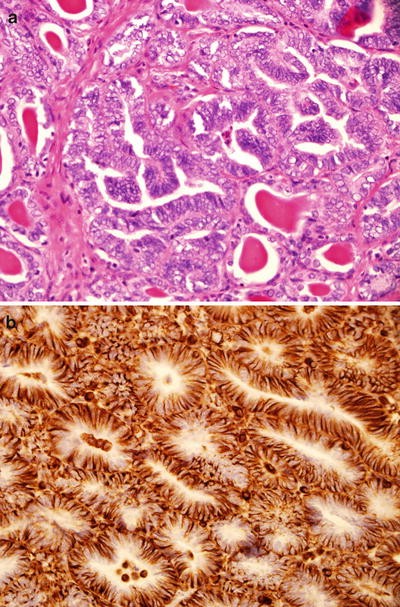
Mesonephric carcinoma on H & E (a) and positive vimentin (b)
-
19.13.
Markers for endometrioid adenocarcinoma of the endometrium (Table 19.13)
Table 19.13 Markers for endometrioid adenocarcinoma of the endometrium Fig. 19.7 
Intense vimentin staining in endometrioid adenocarcinoma
-
19.14.
Markers for serous carcinoma of the endometrium and putative precursors EIC (Table 19.14)
Table 19.14 Markers for serous carcinoma of the endometrium and putative precursors EIC EIC (most commonly used shaded) -
19.15.
Markers for clear cell carcinoma of the endometrium (Table 19.15)
Table 19.15 Markers for clear cell carcinoma of the endometriuma -
19.16.
Markers for carcinosarcoma of the endometrium (Table 19.16)
Table 19.16 Markers for Carcinosarcoma of the endometriuma,b,c -
19.17.
Markers for atypical polypoid adenomyoma (Table 19.17)
Table 19.17 Markers for atypical polypoid adenomyoma (most commonly used shaded) -
19.18.
Markers for stromal nodule and low grade endometrial stromal sarcoma (Table 19.18)
Table 19.18 Markers for stromal nodule and low grade endometrial stromal sarcomaa,b -
19.19.
Markers for high grade endometrial stromal sarcoma (Table 19.19)
Table 19.19 Markers for high grade endometrial stromal sarcomaa,b -
19.20.
Markers for undifferentiated uterine sarcoma (Table 19.20)
Table 19.20 Markers for undifferentiated uterine sarcomaa -
19.21.
Markers for low grade müllerian adenosarcoma (stromal component) (Table 19.21)
Table 19.21 Markers for low grade müllerian adenosarcomaa (stromal component) -
19.22.
Markers for uterine smooth muscle tumors (Table 19.22)
Table 19.22 Markers for uterine smooth muscle tumors -
19.23.
Markers for adenomatoid tumor (Table 19.23)
Table 19.23 Markers for adenomatoid tumor -
19.24.
Markers for PECOMAs (Table 19.24)
Table 19.24 Markers for PECOMAs -
19.25.
Markers for gestational trophoblastic lesions (Table 19.25)
Table 19.25 Markers for gestational trophoblastic lesions -
19.26.
Differentiating high grade SIL of cervix from benign mimics and low grade SIL (Table 19.26)
Table 19.26 Differentiating high grade SIL from benign mimics and low grade SIL -
19.27.
Differentiating in-situ adenocarcinoma of cervix from endometriosis (Table 19.27)
Table 19.27 Differentiating in-situ adenocarcinoma of cervix and endometriosis or tubal-endometrial metaplasia -
19.28.
Differential diagnosis of cervical microglandular hyperplasia (Table 19.28)
Table 19.28 Differential diagnosis of cervical microglandular hyperplasia -
19.29.
Endocervical vs. low grade endometrial adenocarcinoma (Table 19.29)
Table 19.29 Endocervical vs. low grade endometrial adenocarcinomaa -
19.30.
Differentiating Epithelioid Trophoblastic Tumor and cervical squamous cell carcinoma (Table 19.30)
Table 19.30 Differentiating epithelioid trophoblastic tumor and cervical squamous cell carcinoma (most commonly used shaded) -
19.31.
Differentiating adenoid cystic, adenoid basal, basaloid squamous cell, and small cell neuroendocrine carcinomas (Table 19.31)
Table 19.31 Differentiating adenoid cystic (ACC), adenoid basal (ABC), basaloid squamous cell (BSCC), and small cell neuroendocrine (SCNEC) carcinomasa -
19.32.
Arias-Stella reaction and clear cell carcinoma of endometrium (Table 19.32)
Table 19.32 Arias-Stella reaction and clear cell carcinoma of endometriuma -
19.33.
Endometrial adenocarcinoma and carcinosarcoma (MMMT) (Table 19.33)
Table 19.33 Endometrial adenocarcinoma and carcinosarcoma (MMMT)a -
19.34.
Clear cell carcinoma vs malignant mimics with clear cytoplasm (glycogen rich squamous cell carcinoma, clear cell sarcoma, metastatic renal cell carcinoma and yolk sac tumor) (Table 19.34)
Table 19.34 Clear cell carcinoma (CCC) vs. malignant mimics with clear cytoplasm [glycogen rich squamous cell carcinoma (GRSCC), clear cell sarcoma (CCS), metastatic renal cell carcinoma (MCCRCC) and yolk sac tumor (YST)] -
19.35.
Endometrial serous vs endometrioid and clear cell adenocarcinoma (Table 19.35)
Table 19.35 Endometrial serous vs endometrioid and clear cell adenocarcinoma -
19.36.
Useful markers in the differential diagnosis of endometrial undifferentiated carcinoma (Table 19.36)
Table 19.36 Useful markers in the differential diagnosis of endometrial undifferentiated carcinoma (UC) -
19.37.
Differentiating leiomyosarcoma and endometrial stromal sarcoma (Table 19.37)
Table 19.37 Differentiating leiomyosarcoma and endometrial stromal sarcoma (ESS) -
19.38.
Differentiating Leiomyosarcoma and PEComa (Table 19.38)
Table 19.38 Differentiating leiomyosarcoma and PEComa -
19.39.
Differentiating leiomyosarcoma, gastrointestinal stromal tumor, inflammatory myofibroblastic tumor, and spindle cell rhabdomyosarcoma (Table 19.39)
Table 19.39 Differentiating leiomyosarcoma (LMS), gastrointestinal stromal tumor (GIST), inflammatory myofibroblastic tumor (IMT), and spindle cell rhabdomyosarcoma (RHABDO) -
19.40.
Complete hydatidiform mole and partial hydatidiform mole (Table 19.40)
Table 19.40 Complete hydatidiform and partial hydatidiform mole -
19.41.
Epithelioid trophoblastic tumor and poorly differentiated endometrial adenocarcinoma (Table 19.41)
Table 19.41 Epithelioid trophoblastic tumor and poorly differentiated endometrial adenocarcinoma -
19.42.
Differentiating placental site trophoblastic tumors and mimics (exaggerated placental site, epithelioid trophoblastic tumor, choriocarcinoma, epithelioid smooth muscle tumor, and metastatic carcinoma) (Table 19.42)
Table 19.42 Differentiating placental site trophoblastic tumor and mimics [exaggerated placental site (EPS), epithelioid trophoblastic tumor (ETT), choriocarcinoma (CC), epithelioid smooth muscle tumor (ESMT), metastatic carcinoma (MC) and Malignant Melanoma (MM)] Fig. 19.8 
Exaggerated placental site on H & E (a) with negative Ki-67 (b) compared to Choriocarcinoma on H & E (c) that demonstrates extremely elevated Ki-67 proliferative indes (d)
-
19.43.
Summary of common markers of primary uterine carcinoma and the more common metastatic carcinomas (Table 19.43)
Table 19.43 Summary of common markers of primary uterine carcinoma and the more common metastatic carcinomas (recommended markers highlighted)


Note for All Tables
Note: “+”, usually greater than 70 % of cases are positive; “−”, less than 5 % of cases are positive; “+ or −”, usually more than 50 % of cases are positive; “− or +”, less than 50 % of cases are positive. ND no data available, V variable.
References
Rabban JT, Longacre TA. Immunohistology of the female genital tract. In: Dabbs DG, editor. Diagnostic immunohistochemistry – theranostic and genomic applications. 4th ed. Philadelphia, PA: Elsevier Saunders; 2014. p. 653–709.
Fuehrer NE, Keeney GL, Ketterling RP, et al. ALK-1 protein expression and ALK gene rearrangements aid in the diagnosis of inflammatory myofibroblastic tumors of the female genital tract. Arch Pathol Lab Med. 2012;136(6):623–6.
Rabban JT, Zaloudek CJ, Shekitka KM, Tavassoli FA. Inflammatory myofibroblastic tumor of the uterus: a clinicopathologic study of 6 cases emphasizing distinction from aggressive mesenchymal tumors. Am J Surg Pathol. 2005;29(10):1348–55.
Rose PG, Cibas ES, Rose PG, Peters III WA. Cervical squamous neoplasia. In: Crum CP, Nucci MR, Lee KR, editors. Diagnostic gynecologic and obstetric pathology. 2nd ed. Philadelphia, PA: Elsevier Saunders; 2011. p. 245–327.
McCluggage WG. Immunohistochemical and functional biomarkers of value in female genital tract lesions. In: Robboy SJ, Mutter GL, Prat J, Bentley R, Russell P, Anderson MC, editors. Robboy’s pathology of the female reproductive tract. 2nd ed. London: Churchill Livingstone; 2009. p. 999–1010.
O'Neill CJ, McCluggage WG. p16 expression in the female genital tract and its value in diagnosis. Adv Anat Pathol. 2006;13(1):8–15.
Herfs M, Yamamoto Y, Laury A, et al. A discrete population of squamocolumnar junction cells implicated in the pathogenesis of cervical cancer. Proc Natl Acad Sci USA. 2012;109(26):10516–21.
Pinto AP, Schlecht NF, Woo TY, et al. Biomarker (ProEx C, p16(INK4A), and MiB-1) distinction of high-grade squamous intraepithelial lesion from its mimics. Mod Pathol. 2008;21(9):1067–74.
Kong CS, Balzer BL, Troxell ML, et al. p16INK4A immunohistochemistry is superior to HPV in situ hybridization for the detection of high-risk HPV in atypical squamous metaplasia. Am J Surg Pathol. 2007;31(1):33–43.
Herfs M, Parra-Herran C, Howitt BE, et al. Cervical squamocolumnar junction-specific markers define distinct, clinically relevant subsets of low-grade squamous intraepithelial lesions. Am J Surg Pathol. 2013;37(9):1311–8.
Herfs M, Crum CP. Laboratory management of cervical intraepithelial neoplasia: proposing a new paradigm. Adv Anat Pathol. 2013;20(2):86–94.
Singer G, Kurman RJ, McMaster MT, et al. HLA-G immunoreactivity is specific for intermediate trophoblast in gestational trophoblastic disease and can serve as a useful marker in differential diagnosis. Am J Surg Pathol. 2002;26(7):914–20.
Ou-Yang RJ, Hui P, Yang XJ, Zynger DL. Expression of glypican 3 in placental site trophoblastic tumor. Diagn Pathol. 2010;5(1):64.
Kindelberger DW, Krane JE, Lee KR. Glandular neoplasia of the cervix. In: Crum CP, Nucci MR, Lee KR, editors. Diagnostic gynecologic and obstetric pathology. 2nd ed. Philadelphia, PA: Elsevier Saunders; 2011. p. 328–78.
Negri G, Egarter-Vigl E, Kasal A, et al. p16INK4a is a useful marker for the diagnosis of adenocarcinoma of the cervix uteri and its precursors: An immunohistochemical study with immunocytochemical correlations. Am J Surg Pathol. 2003;27(2):187–93.
McCluggage WG. Immunohistochemistry as a diagnostic aid in cervical pathology. Pathology. 2007;39(1):97–111.
Kong CS, Beck AH, Longacre TA. A panel of 3 markers including p16, ProExC, or HPV ISH is optimal for distinguishing between primary endometrial and endocervical adenocarcinomas. Am J Surg Pathol. 2010;34(7):915–26.
McCluggage WG. Ten problematic issues identified by pathology review for multidisciplinary gynaecological oncology meetings. J Clin Pathol. 2012;65(4):293–301.
DeLair D, Soslow R, Gilks B, et al. The morphologic spectrum of immunohistochemically characterized clear cell carcinoma of the ovary: a study of 83 cases. Mod Pathol. 2009;22 Suppl 1:211A.
Fadare O, Liang SX. Diagnostic utility of hepatocyte nuclear factor 1-beta immunoreactivity in endometrial carcinomas: lack of specificity for endometrial clear cell carcinoma. Appl Immunohistochem Mol Morphol. 2012;20(6):580–7.
Tsuchiya A, Sakamoto M, Yasuda J, et al. Expression profiling in ovarian clear cell carcinoma: identification of hepatocyte nuclear factor-1 beta as a molecular marker and a possible molecular target for therapy of ovarian clear cell carcinoma. Am J Pathol. 2003;163(6):2503–12.
Kato N, Sasou S, Motoyama T. Expression of hepatocyte nuclear factor-1beta (HNF-1beta) in clear cell tumors and endometriosis of the ovary. Mod Pathol. 2006;19(1):83–9.
Yamamoto S, Tsuda H, Aida S, et al. Immunohistochemical detection of hepatocyte nuclear factor 1beta in ovarian and endometrial clear-cell adenocarcinomas and nonneoplastic endometrium. Hum Pathol. 2007;38(7):1074–80.
Kobel M, Kalloger SE, Carrick J, et al. A limited panel of immune-markers can reliably distinguish between clear cell and high-grade serous carcinoma of the ovary. Am J Surg Pathol. 2009;33(1):14–21.
Hoang LN, Han G, McConechy M, et al. Immunohistochemical characterization of prototypical endometrial clear cell carcinoma – diagnostic utility of HNF-1b and oestrogen receptor. Histopathology. 2014;64(4):585–96.
McConechy MK, Ding J, Cheang MC, et al. Use of mutation profiles to refine the classification of endometrial carcinomas. J Pathol. 2012;228(1):20–30.
Al-Loh S, Al-Hussaini M. Undifferentiated endometrial carcinoma: a diagnosis frequently overlooked. Arch Pathol Lab Med. 2013;137(3):438–42.
Altrabulsi B, Malpica A, Deavers MT, et al. Undifferentiated carcinoma of the endometrium. Am J Surg Pathol. 2005;29(10):1316–21.
Silva EG, Deavers MT, Malpica A. Undifferentiated carcinoma of the endometrium: a review. Pathology. 2007;39(1):134–8.
Silva EG, Deavers MT, Bodurka DC, et al. Association of low grade endometrioid carcinoma of the uterus and ovary with undifferentiated carcinoma: a new type of dedifferentiated carcinoma? Int J Gynecol Pathol. 2006;25(1):52–8.
Soslow RA. Endometrial carcinomas with ambiguous features. Semin Diagn Pathol. 2010;27(4):261–73.
McCluggage WG. New developments in endocervical glandular lesions. Histopathology. 2013;62(1):138–60.
Yemelyanova A, Ji H, Shih I, et al. Utility of p16 expression for distinction of uterine serous carcinomas from endometrial endometrioid and endocervical adenocarcinomas: immunohistochemical analysis of 201 cases. Am J Surg Pathol. 2009;33(10):1504–14.
Chiesa-Vottero AG, Malpica A, Deavers MT, Broaddus R, Nuovo GJ, Silva EG. Immunohistochemical overexpression of p16 and p53 in uterine serous carcinoma and ovarian high-grade serous carcinoma. Int J Gynecol Pathol. 2007;26(3):328–33.
Saad RS, Mashhour M, Noftech-Moses S, et al. P16INK4a expression in undifferentiated carcinoma of the uterus does not exclude its endometrial origin. Int J Gynecol Pathol. 2012;31(1):57–65.
Hirschowitz L, Ganesan R, McCluggage WG. WT1, p53 and hormone receptor expression in uterine serous carcinoma. Histopathology. 2009;55(4):478–82.
McCluggage WG, Soslow RA, Gilks CB. Patterns of p53 immunoreactivity in endometrial carcinomas: ‘all or nothing’ staining is of importance. Histopathology. 2011;59(4):786–8.
Mutter GL, Ince TA, Baak JP, Kust GA, Zhou XP, Eng C. Molecular identification of latent precancers in histologically normal endometrium. Cancer Res. 2001;61(11):4311–4.
Mutter GL, Lin MC, Fitzgerald JT, et al. Altered PTEN expression as a diagnostic marker for the earliest endometrial precancers. J Natl Cancer Inst. 2000;92(11):924–30.
Latta E, Chapman WB. PTEN mutations and evolving concepts in endometrial neoplasia. Curr Opin Obstet Gynecol. 2002;14(1):59–65.
Mutter GL, Duska LR, Crum CP. Preinvasive endometrial neoplasia. In: Crum CP, Nucci MR, Lee KR, editors. Diagnostic gynecologic and obstetric pathology. 2nd ed. Philadelphia, PA: Elsevier Saunders; 2011. p. 457–89.
Park KJ, Bramlage MP, Ellenson LH, Pirog EC. Immunoprofile of adenocarcinomas of the endometrium, endocervix, and ovary with mucinous differentiation. Appl Immunohistochem Mol Morphol. 2009;17(1):8–11.
Quade BJ, Yang A, Wang Y, et al. Expression of the p53 homo-logue p63 in early cervical neoplasia. Gynecol Oncol. 2001;80(1):24–9.
Ip PP, Irving JA, McCluggage WG, et al. Papillary proliferation of the endometrium: a clinicopathologic study of 59 cases of simple and complex papillae without cytologic atypia. Am J Surg Pathol. 2013;37(2):167–77.
Riethdorf L, Riethdorf S, Lee KR, Cviko A, Loning T, Crum CP. Human papillomaviruses, expression of p16, and early endocervical glandular neoplasia. Hum Pathol. 2002;33(9):899–904.
Gilks CB, Young RH, Aguirre P, DeLellis RA, Scully RE. Adenoma malignum (minimal deviation adenocarcinoma) of the uterine cervix. A clinicopathological and immunohistochemical analysis of 26 cases. Am J Surg Pathol. 1989;13(9):717–29.
Mikami Y, Kiyokawa T, Moriya T, Sasano H. Immunophenotypic alteration of the stromal component in minimal deviation adenocar- cinoma (“adenoma malignum”) and endocervical glandular hyper-plasia: a study using oestrogen receptor and alpha-smooth muscle actin double immunostaining. Histopathology. 2005;46(2):130–6.
Utsugi K, Hirai Y, Takeshima N, Akiyama F, Sakurai S, Hasumi K. Utility of the monoclonal antibody HIK1083 in the diagnosis of adenoma malignum of the uterine cervix. Gynecol Oncol. 1999;75(3):345–8.
Park KJ, Kiyokawa T, Soslow RA, et al. Unusual endocervical adenocarcinomas: an immunohistochemical analysis with molecular detection of human papillomavirus. Am J Surg Pathol. 2011;35(5):633–46.
Kusanagi Y, Kojima A, Mikami Y, et al. Absence of high-risk human papillomavirus (HPV) detection in endocervical adenocarcinoma with gastric morphology and phenotype. Am J Pathol. 2010;177(5):2169–75.
Rabban JT, McAlhany S, Lerwill MF, Grenert JP, Zaloudek CJ. PAX2 distinguishes benign mesonephric and mullerian glandular lesions of the cervix from endocervical adenocarcinoma, including minimal deviation adenocarcinoma. Am J Surg Pathol. 2010;34(2):137–46.
Emanuel P, Wang B, Wu M, Burstein DE. p63 Immunohistochemistry in the distinction of adenoid cystic carcinoma from basaloid squamous cell carcinoma. Mod Pathol. 2005;18(5):645–50.
Gardner GJ, Reidy-Lagunes D, Gehrig PA. Neuroendocrine tumors of the gynecologic tract: A Society of Gynecologic Oncology (SGO) clinical document. Gynecol Oncol. 2011;122(1):190–8.
Li JD, Zhuang Y, Li YF, et al. A clinicopathological aspect of primary small-cell carcinoma of the uterine cervix: a single-centre study of 25 cases. J Clin Pathol. 2011;64(12):1102–7.
Houghton O, McCluggage WG. The expression and diagnostic utility of p63 in the female genital tract. Adv Anat Pathol. 2009;16(5):316–21.
Young RH, Clement PB. Endocervical adenocarcinoma and its variants: their morphology and differential diagnosis. Histopathology. 2002;41(3):185–207.
Ordi J, Nogales FF, Palacin A, et al. Mesonephric adenocarcinoma of the uterine corpus: CD10 expression as evidence of mesonephric differentiation. Am J Surg Pathol. 2001;25(12):1540–5.
Ordi J, Romagosa C, Tavassoli FA, et al. CD10 expression in epithelial tissues and tumors of the gynecologic tract: a useful marker in the diagnosis of mesonephric, trophoblastic, and clear cell tumors. Am J Surg Pathol. 2003;27(2):178–86.
Silver SA, Devouassoux-Shisheboran M, Mezzetti TP, Tavassoli FA. Mesonephric adenocarcinomas of the uterine cervix: a study of 11 cases with immunohistochemical findings. Am J Surg Pathol. 2001;25(3):379–87.
Gilks CB, Oliva E, Soslow R. Poor interobserver reproducibility in the diagnosis of high-grade endometrial carcinoma. Am J Surg Pathol. 2013;37(6):874–81.
Soslow R. High-grade endometrial carcinomas – strategies for typing. Histopathology. 2013;62(1):89–110.
Darvishian F, Hummer AJ, Thaler HT, et al. Serous endometrial cancers that mimic endometrioid adenocarcinomas: a clinicopathologic and immunohistochemical study of a group of problematic cases. Am J Surg Pathol. 2004;28(12):1568–78.
Lomo L, Nucci MR, Lee KR, et al. Histologic and immunohistochemical decision making in endometrial adenocarcinoma. Mod Pathol. 2008;21(8):937–42.
Reid-Nicholson M, Iyengar P, Hummer AJ, et al. Immunophenotypic diversity of endometrial adenocarcinomas: implications for differential diagnosis. Mod Pathol. 2006;19(8):1091–100.
Alkushi A, Clarke BA, Akbari M, et al. Identification of prognostically relevant and reproducible subsets of endometrial adenocarcinoma based on clustering analysis of immunostaining data. Mod Pathol. 2007;20(11):1156–65.
Risinger JL, Hayes K, Maxwell GL. PTEN mutation in endometrial cancers is associated with favorable clinical and pathologic characteristics. Clin Cancer Res. 1998;4(12):3005–10.
Fukuchi T, Sakamoto M, Tsuda H, Maruyama K, Nozawa S, Hirohashi S. Beta-catenin mutation in carcinoma of the uterine endometrium. Cancer Res. 1998;58(16):3526–8.
Goldstein NS, Uzieblo A. WT1 immunoreactivity in uterine papillary serous carcinomas is different from ovarian serous carcinomas. Am J Clin Pathol. 2002;117(4):541–5.
Lax SF, Kendall B, Tashiro H, Slebos RJ, Hedrick L. The frequency of p53, K-ras mutations, and microsatellite instability differs in uterine endometrioid and serous carcinoma: evidence of distinct molecular genetic pathways. Cancer. 2000;88(4):814–24.
Pallares J, Bussaglia E, Martinez-Guitarte JL, et al. Immunohistochemical analysis of PTEN in endometrial carcinoma: a tissue microarray study with a comparison of four commercial antibodies in correlation with molecular abnormalities. Mod Pathol. 2005;18(5):719–27.
Peiro G, Diebold J, Lohse P, et al. Microsatellite instability, loss of heterozygosity, and loss of hMLH1 and hMSH2 protein expression in endometrial carcinoma. Hum Pathol. 2002;33(3):347–54.
Schlosshauer PW, Ellenson LH, Soslow RA. Beta-catenin and E-cadherin expression patterns in high-grade endometrial carcinoma are associated with histological subtype. Mod Pathol. 2002;15(10):1032–7.
Quick CM, Laury AR, Monte NM, Mutter GL. Utility of PAX2 as a Marker for Diagnosis of Endometrial Intraepithelial Neoplasia. Am J Clin Pathol. 2012;138(5):678–84.
Liang SX, Chambers SK, Cheng L, et al. Endometrial glandular dysplasia: A putative precursor lesion of uterine papillary serous carcinoma. Part II: molecular features. Int J Surg Pathol. 2004;12(4):319–31.
Zheng W, Liang SX, Yu H, et al. Endometrial glandular dysplasia: a newly defined precursor lesion of uterine papillary serous carcinoma. Part I: morphologic features. Int J Surg Pathol. 2004;12(3):207–23.
Alkushi A, Kobel M, Kalloger SE, Gilks CB. High-grade endometrial carcinoma: serous and grade 3 endometrioid carcinomas have different immunophenotypes and outcomes. Int J Gynecol Pathol. 2010;29(4):343–50.
DeLair D, Soslow RA. Endometrial clear cell carcinomas with and without aberrant p53 expression: a study of 16 cases. Lab Invest. 2012;92:265–266A.
Mikami Y, Hata S, Kiyokawa T, Manabe T. Expression of CD10 in malignant mullerian mixed tumors and adenosarcomas: an immunohistochemical study. Mod Pathol. 2002;15(9):923–30.
Kurihara S, Oda Y, Ohishi Y, et al. Endometrial stromal sarcomas and related high-grade sarcomas: immunohistochemical and molecular genetic study of 31 cases. Am J Surg Pathol. 2008;32(8):1228–38.
Jung CK, Jung JH, Lee A, et al. Diagnostic use of nuclear beta-catenin expression for the assessment of endometrial stromal tumors. Mod Pathol. 2008;6:756–63.
McCluggage WG, Sumathi VP, Maxwell P. CD10 is a sensitive and diagnostically useful immunohistochemical marker of normal endometrial stroma and of endometrial stromal neoplasms. Histopathology. 2001;39(3):273–8.
Oliva E, Young RH, Clement PB, Bhan AK, Scully RE. Cellular benign mesenchymal tumors of the uterus. A comparative morphologic and immunohistochemical analysis of 33 highly cellular leiomyomas and six endometrial stromal nodules, two frequently confused tumors. Am J Surg Pathol. 1995;19(7):757–68.
Sumathi VP, Al-Hussaini M, Connolly LE, et al. Endometrial stromal neoplasms are immunoreactive with WT-1 antibody. Int J Gynecol Pathol. 2004;23(3):241–7.
Yilmaz A, Rush DS, Soslow RA. Endometrial stromal sarcomas with unusual histologic features: a report of 24 primary and metastatic tumors emphasizing fibroblastic and smooth muscle differentiation. Am J Surg Pathol. 2002;26(9):1142–50.
Kurihara S, Oda Y, Ohishi Y, et al. Coincident expression of beta-catenin and cyclin- D1 in endometrial stromal tumors and related high-grade sarcomas. Mod Pathol. 2010;23(2):225–34.
Croce S, Hostein I, Ribeiro A, et al. YWHAE rearrangement identified by FISH and RT-PCR in endometrial stromal sarcomas: genetic and pathological correlations. Mod Pathol. 2013;26(10):1390–400.
Lee CH, Ali RH, Rouzbahman M, et al. Cyclin D1 as a diagnostic immunomarker for endometrial stromal sarcoma with YWHAE-FAM22 rearrangement. Am J Surg Pathol. 2012;36(10):1562–70.
Amant F, Steenkiste E, Schurmans K, et al. Immunohistochemical expression of CD10 antigen in uterine adenosarcoma. Int J Gynecol Cancer. 2004;14(6):1118–21.
Soslow RA, Ali A, Oliva E. Mullerian adenosarcomas: an immunophenotypic analysis of 35 cases. Am J Surg Pathol. 2008;32(7):1013–21.
Gannon BR, Manduch M, Childs TJ. Differential immunoreactivity of p16 in leiomyosarcomas and leiomyoma variants. Int J Gynecol Pathol. 2008;27(1):68–73.
Mittal K, Demopoulos RI. MIB-1 (Ki-67), p53, estrogen receptor, and progesterone receptor expression in uterine smooth muscle tumors. Hum Pathol. 2001;32(9):984–7.
Oliva E, Clement PB, Young RH. Epithelioid endometrial and endometrioid stromal tumors: a report of four cases emphasizing their distinction from epithelioid smooth muscle tumors and other oxyphilic uterine and extrauterine tumors. Int J Gynecol Pathol. 2002;21(1):48–55.
Silva EG, Deavers MT, Bodurka DC, Malpica A. Uterine epithelioid leiomyosarcomas with clear cells: reactivity with HMB-45 and the concept of PEComa. Am J Surg Pathol. 2004;28(2):244–9.
Winter 3rd WE, Seidman JD, Krivak TC, et al. Clinicopathological analysis of c-kit expression in carcinosarcomas and leiomyosarcomas of the uterine corpus. Gynecol Oncol. 2003;91(1):3–8.
Iwata J, Fletcher CD. Immunohistochemical detection of cytokeratin and epithelial membrane antigen in leiomyosarcoma: a systematic study of 100 cases. Pathol Int. 2000;50(1):7–14.
Fukunaga M, Nomura K, Endo Y, et al. Carcinosarcoma of the uterus with extensive neuroectodermal differentiation. Histopathology. 1996;29(6):565–70.
Kanamori T, Takakura K, Mandai M, Kariya M, Fukuhara K, Sakaguchi M, et al. Increased expression of calcium-binding protein S100 in human uterine smooth muscle tumours. Mol Hum Reprod. 2004;10(10):735–42.
Carvalho JC, Thomas DG, Lucas DR. Cluster analysis of immunohistochemical markers in leiomyosarcoma delineates specific anatomic and gender subgroups. Cancer. 2009;115(18):4186–95.
Lee CH, Turbin DA, Sung YC, et al. A panel of antibodies to determine site of origin and malignancy in smooth muscle tumors. Mod Pathol. 2009;22(12):1519–31.
O'Neill CJ, McBride HA, Connolly LE, McCluggage WG. Uterine leiomyosarcomas are characterized by high p16, p53 and MIB1 expression in comparison with usual leiomyomas, leiomyoma variants and smooth muscle tumours of uncertain malignant potential. Histopathology. 2007;50(7):851–8.
D’Angelo E, Espinosa I, Ali R, et al. Uterine leiomyosarcomas: tumor size, mitotic index, and biomarkers ki67, and bcl-2 identify two groups with different prognosis. Gynecol Oncol. 2011;121(2):328–33.
Hakverdi S, Gungoren A, Yaldiz M, Hakverdi AU, Toprak S. Immunohistochemical analysis of p16 expression in uterine smooth muscle tumors. Eur J Gynaecol Oncol. 2011;32(5):513–5.
Ip PP, Cheung AN, Clement PB. Uterine smooth muscle tumors of uncertain malignant potential (stump): a clinicopathologic analysis of 16 cases. Am J Surg Pathol. 2009;33(7):992–1005.
Akhan SE, Yavuz E, Tecer A, et al. The expression of ki-67, p53, estrogen and progesterone receptors affecting survival in uterine leiomyosarcomas. A clinicopathologic study. Gynecol Oncol. 2005;99(1):36–42.
Chen L, Yang B. Immunohistochemical analysis of p16, p53, and ki-67 expression in uterine smooth muscle tumors. Int J Gynecol Pathol. 2008;27(3):326–32.
Kefeli M, Yildiz L, Kaya FC, et al. Fascin expression in uterine smooth muscle tumors. Int J Gynecol Pathol. 2009;28(4):328–33.
Otis CN. Uterine adenomatoid tumors: immunohistochemical characteristics with emphasis on Ber-EP4 immunoreactivity and distinction from adenocarcinoma. Int J Gynecol Pathol. 1996;15(2):146–51.
Schwartz EJ, Longacre TA. Adenomatoid tumors of the female and male genital tracts express WT1. Int J Gynecol Pathol. 2004;23(2):123–8.
Fadare O. Perivascular epithelioid cell tumors (PEComas) and smooth muscle tumors of the uterus. Am J Surg Pathol. 2007;31(9):1454–5.
Ye HY, Chen JG, Luo DL, Jiang ZM, Chen ZH. Perivascular epithelioid cell tumor (PEComa) of gynecologic origin: a clinicopathological study of three cases. Eur J Gynaecol Oncol. 2012;33(1):105–8.
Wu JH, Zhou JL, Cui Y, et al. Malignant perivascular epithelioid cell tumor of the retroperitoneum. Int J Clin Exp Pathol. 2013;6(10):2251–6.
Folpe AL, Mentzel T, Lehr HA, et al. Perivascular epithelioid cell neoplasms of soft tissue and gynecologic origin: a clinicopathologic study of 26 cases and review of the literature. Am J Surg Pathol. 2005;29(12):1558–75.
Vang R, Kempson RL. Perivascular epithelioid cell tumor (PEComa) of the uterus: a subset of HMB-45-positive epithelioid mesenchymal neoplasms with an uncertain relationship to pure smooth muscle tumors. Am J Surg Pathol. 2002;26(1):1–13.
Fisher RA, Hodges MD, Rees HC, et al. The maternally tran-scribed gene p57(KIP2) (CDNK1C) is abnormally expressed in both androgenetic and biparental complete hydatidiform moles. Hum Mol Genet. 2002;11(26):3267–72.
Jun SY, Ro JY, Kim KR. p57kip2 is useful in the classification and differential diagnosis of complete and partial hydatidiform moles. Histopathology. 2003;43(1):17–25.
Sebire NJ, Rees HC, Peston D, Seckl MJ, Newlands ES, Fisher RA. p57(KIP2) immunohistochemical staining of gestational tro-phoblastic tumours does not identify the type of the causative pregnancy. Histopathology. 2004;45(2):135–41.
Shih IM, Kurman RJ. Immunohistochemical localization of inhibin-alpha in the placenta and gestational trophoblastic lesions. Int J Gynecol Pathol. 1999;18(2):144–50.
Kommoss F, Schmidt D, Coerdt W, et al. Immunohistochemical expression analysis of inhibin-alpha and -beta subunits in partial and complete moles, trophoblastic tumors, and endometrial decidua. Int J Gynecol Pathol. 2001;20(4):380–5.
Shih IM, Kurman RJ. Ki-67 labeling index in the differential diagnosis of exaggerated placental site, placental site trophoblastic tumor, and choriocarcinoma: a double immunohistochemical staining technique using Ki-67 and Mel-CAM antibodies. Hum Pathol. 1998;29(1):27–33.
Wong SC, Chan AT, Chan JK, Lo YM. Nuclear beta-catenin and Ki-67 expression in choriocarcinoma and its pre-malignant form. J Clin Pathol. 2006;59(4):387–92.
Shih I. Trophogram, an immunohistochemistry-based algorithmic approach, in the differential diagnosis of trophoblastic tumors and tumorlike lesions. Ann Diagn Pathol. 2007;11(3):228–34.
Kalhor N, Ramirez PT, Deavers MT, et al. Immunohistochemical studies of trophoblastic tumors. Am J Surg Pathol. 2009;33(4):633–8.
Lee Y, Kim KR, McKeon F, et al. A unifying concept of trophoblastic differentiation and malignancy defined by biomarker expression. Hum Pathol. 2007;38(7):1003–13.
Shih IM, Kurman RJ. p63 Expression is useful in the distinction of epithelioid trophoblastic and placental site trophoblastic tumors by profiling trophoblastic subpopulations. Am J Surg Pathol. 2004;28(9):1177–83.
Albores-Saavedra J, Latif S, Carrick KS, Alvarado-Cabrero I, Fowler MR. CD56 reactivity in small cell carcinoma of the uterine cervix. Int J Gynecol Pathol. 2005;24(2):113–7.
Wang HL, Lu DW. Detection of human papillomavirus DNA and expression of p16, Rb, and p53 proteins in small cell carcinomas of the uterine cervix. Am J Surg Pathol. 2004;28(7):901–8.
Grayson W, Taylor LF, Cooper K. Adenoid cystic and adenoid basal carcinoma of the uterine cervix: comparative morphologic, mucin, and immunohistochemical profile of two rare neoplasms of putative ‘reserve cell’ origin. Am J Surg Pathol. 1999;23(4):448–58.
Vang R, Whitaker BP, Farhood AI, Silva EG, Ro JY, Deavers MT. Immunohistochemical analysis of clear cell carcinoma of the gynecologic tract. Int J Gynecol Pathol. 2001;20(3):252–9.
Vang R, Barner R, Wheeler DT, Strauss BL. Immunohistochemical staining for Ki-67 and p53 helps distinguish endometrial Arias-Stella reaction from high-grade carcinoma, including clear cell carcinoma. Int J Gynecol Pathol. 2004;23(3):223–33.
McCluggage WG. Morphological subtypes of ovarian carcinoma: a review with emphasis on new developments and pathogenesis. Pathology. 2011;43(5):420–32.
Mhawech-Fauceglia P, Yan L, Liu S, Pejovic T. ER+/PR+/TFF3+/IMP3_ immunoprofile distinguishes endometrioid from serous and clear cell carcinomas of the endometrium: a study of 401 cases. Histopathology. 2013;62:976–85.
Author information
Authors and Affiliations
Corresponding author
Editor information
Editors and Affiliations
Rights and permissions
Copyright information
© 2015 Springer Science+Business Media New York
About this chapter
Cite this chapter
Kaspar, H.G., Crum, C.P. (2015). Uterus. In: Lin, F., Prichard, J. (eds) Handbook of Practical Immunohistochemistry. Springer, New York, NY. https://doi.org/10.1007/978-1-4939-1578-1_19
Download citation
DOI: https://doi.org/10.1007/978-1-4939-1578-1_19
Published:
Publisher Name: Springer, New York, NY
Print ISBN: 978-1-4939-1577-4
Online ISBN: 978-1-4939-1578-1
eBook Packages: MedicineMedicine (R0)