
Abstract
Acute aortic conditions include, but are not limited to, aortic rupture, aortic dissection, intramural hematoma, and penetrating aortic ulcer. Prompt diagnosis of these conditions is essential for managing these conditions. Because these conditions often have similar symptoms, namely, chest and abdominal pain, the imaging characteristics are key to prompt and accurate diagnosis.
Access provided by Autonomous University of Puebla. Download chapter PDF
Keywords
These keywords were added by machine and not by the authors. This process is experimental and the keywords may be updated as the learning algorithm improves.
Introduction
Acute aortic conditions include, but are not limited to, aortic rupture, aortic dissection, intramural hematoma, and penetrating aortic ulcer. Prompt diagnosis of these conditions is essential for managing these conditions. Because these conditions often have similar symptoms, namely, chest and abdominal pain, the imaging characteristics are key to prompt and accurate diagnosis.
Abdominal Aortic Aneurysm and Aortic Rupture
Abdominal aortic aneurysm (AAA) is seen in 5–10 % of elderly male smokers. Most AAAs are true aneurysms and involve all three layers of the aortic wall. The two most common etiologies of AAA are degenerative and inflammatory (Table 1.1).
Other less frequent etiologies of AAA include mycotic aneurysm, which constitutes 1–3 % of aortic aneurysms. However, mycotic aneurysm is known to more commonly involve aorta than any other artery. Staphylococcus and Streptococcus species are the most common pathogens of mycotic aneurysm. The cases of mycotic aneurysm due to Salmonella species are more common in East Asia and demonstrate an early tendency to rupture.
The most significant complication of AAA is aortic rupture. The mortality rate for ruptured AAA is 50 %; thus, an accurate diagnosis is essential for prompt surgical intervention. The risk of rupture is proportional to the maximum cross-sectional diameter, with 1 %/year risk for aneurysms measuring 5–5.9 cm. The risk of rupture increases up to 20 %/year for an aneurysm measuring greater than 7 cm in diameter. Although AAAs are less common in females (M:F = 4:1), they are more likely to rupture when compared to males.
Ultrasound is the most commonly used imaging modality to screen for AAA and has been shown to reduce mortality. The imaging criteria to diagnose AAA include aortic caliber of more than 3 cm and an aortic caliber of more than 1.5 times the expected diameter of the abdominal aorta (Fig. 1.1). The aortic caliber is measured perpendicular to the long axis of the aorta, from outer wall to outer wall. Although ultrasound is highly sensitive in making the diagnosis of abdominal aortic aneurysm, it is not as reliable as CT in diagnosing aortic rupture. However, the demonstration of normal caliber of abdominal aorta by ultrasound makes aortic rupture an unlikely possibility.

Saccular abdominal aortic aneurysm. (a and b) US demonstrate a saccular infrarenal aortic aneurysm (curved arrow) with yin-yang sign on color Doppler imaging. (c) Sagittal reformation demonstrates the saccular infrarenal abdominal aortic aneurysm (curved arrow)
Most aortic aneurysms rupture involves the middle third of the aneurysm, through the posterolateral wall and into the retroperitoneum (Fig. 1.2a). However, intraperitoneal rupture and rupture into the bowel (usually the duodenum) and very rarely into the IVC may occur (Fig. 1.2b, c).

Abdominal aortic aneurysm rupture, aorto-enteric and aortocaval fistula. (a) Contrast-enhanced CT scan study of the lower abdomen demonstrates active extravasation of contrast (arrow) from infrarenal abdominal aortic aneurysm. There is retroperitoneal hemorrhage (arrowheads) identified around the aortic aneurysm. (b) Aorto-enteric fistula. Contrast-enhanced CT scan study demonstrates communication (arrowhead) of the third portion of the duodenum (arrow) with the infrarenal abdominal aortic aneurysm sac. The patient had recently undergone endovascular stent placement. (c) Aortocaval fistula. Doppler US shows the combination of arterial and venous spectral waveform in the inferior vena cava lumen, in a patient with aortocaval fistula
Risk Factors for Aortic Rupture
Progressive aneurysmal dilatation of the aorta with increased wall tension is directly related to the risk of rupture. The decreased proportion of thrombus to lumen ratio is also thought to play a part, as a larger thrombus better protects against rupture by providing protection against the high aortic pressures [1]. In addition, the amount of thrombus calcification, which is thought to be related to the amount of thrombus present, is also an indirect measure [2].
Imaging
The imaging modality of choice is a contrast-enhanced multidetector CT (MDCT). The CT can demonstrate an AAA with surrounding retroperitoneal hemorrhage into psoas compartment, pararenal space, and perirenal space. A contrast-enhanced CT provides additional information about the aortic size, presence or absence of active extravasation, and anatomic relationships (Table 1.2). A hyperdense crescent sign and draped aorta sign are indicators of contained aortic leak or impending rupture. Focal discontinuity of intimal calcification is also a secondary sign of aortic rupture.
Hyperdense Crescent Sign
Hyperdense crescent sign is seen as a well-defined peripheral, high-density, crescent configuration within a thrombus where there is internal dissection of hemorrhage into the thrombus and ultimately reaching the aortic wall. It is a sign of acute or impending rupture (Fig. 1.3a) [1].


CT features of abdominal aortic aneurysm rupture. (a) Hyperdense crescent sign. Noncontrast CT demonstrates large retroperitoneal hematoma (arrowhead) from the ruptured aortic aneurysm. A hyperdense crescent (curved arrow) is present in the anterior wall of the infrarenal abdominal aortic aneurysm. (b) Draped aorta sign. Contrast-enhanced CT demonstrates draping of the posterior wall of the aorta (straight arrows) on the anterior aspect of the lumbar spine. There is large retroperitoneal hematoma (curved arrow) identified in the psoas compartment and left posterior pararenal space. (c) Schematic representation of the draped aorta sign (a) and hyperdense crescent sign (b). Draped aorta sign is characterized by draping of deficient aortic wall on the anterior aspect of the vertebral body. Hyperdense crescent sign is characterized by the presence of a high-density sickle-shaped blood clot in the aortic wall. (d) Tangential calcium sign in a patient with contained aortic leak. Noncontrast CT demonstrates intimal calcifications (arrowheads) displaced from their expected location and pointing away from the aortic circumference. (e) Rupture of mycotic aneurysm. Noncontrast CT demonstrates air in the wall of the aortic aneurysm, secondary to clostridial infection. Breech in the aortic wall is indicated by the presence of air (arrowheads) outside the aortic adventitia
Draped Aorta Sign
Draped aorta sign indicates a contained aortic rupture and shows posterior aortic wall not identifiable as a separate structure and draping over the adjacent vertebral bodies (Fig. 1.3b, c). If rupture should occur, the most common sign of aneurysmal rupture is a retroperitoneal hematoma adjacent to the aneurysm.
Tangential Calcium Sign
The intimal calcification in the aorta points away from the circumference of the aneurysm (Fig. 1.3d).
The typical imaging features of mycotic aneurysm (Fig. 1.3e) include rapidly increasing caliber of a saccular aortic aneurysm with wall irregularity, periaortic edema and soft tissue mass, and the presence of gas. Periaortic soft tissue stranding and soft tissue mass are the most common features seen on imaging of mycotic aneurysm. Calcifications and thrombus are uncommon in a mycotic aneurysm. The lack of calcification in the aortic wall is due to the nonatherosclerotic origin of the aneurysm.
Aortic Dissection
Aortic dissection is the most common acute presentation involving the aorta [3]. It usually originates with a tear in the intima, which causes high-pressure blood to enter and dissect the aortic wall (Table 1.3).
The most commonly used classification for aortic dissection is the Stanford classification system.
-
1.
Type A aortic dissection: Regardless of origin and extent of dissection, a Type A aortic dissection involves the ascending aorta (Fig. 1.4) [4]. The potential for complications with Type A dissection necessitates urgent surgical intervention [4]. The complications include dissection into the pericardium resulting in cardiac tamponade, dissection into the coronary arteries resulting in occlusion, and aortic insufficiency with involvement of the valve [4].
Fig. 1.4 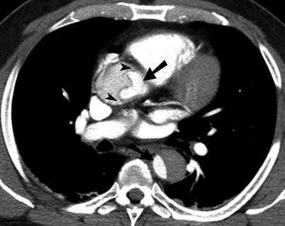
Type A aortic dissection. Contrast-enhanced CT scan study of the chest demonstrates aortic dissection involving the ascending as well as the descending thoracic aorta. The true lumen can be identified by the smaller caliber (arrows) and higher density. The beak sign (arrowheads) is identified in the false lumen
-
2.
Type B aortic dissection: The aortic dissection originates past the left subclavian artery [5]. Unlike Type A dissection, the Type B dissections are usually medically treated.

Imaging
The imaging modality of choice to evaluate aortic dissection is MDCT. It allows accurate assessment of the extent of the disease, including the origin of the dissection, involvement of the visceral branches, and presence of a false lumen [3, 4]. The most characteristic findings of aortic dissection include an intimal flap and two distinct lumens. Secondary findings include intimal displacement of calcified wall, delayed enhancement of false lumen, pericardial or mediastinal hematoma, and ischemia or infarction of distal organs supplied by the false lumen [4].
True Versus False Lumen
Once the recognition of an aortic dissection is made, it is important to distinguish between the true and false lumen for treatment purposes, especially endovascular repair. Lepage et al. evaluated signs to distinguish between the true and false lumen and determined two consistent signs: beak sign and larger cross-sectional area of false lumen as the best indicators. The beak sign is present in the false lumen and consists of an acute angle between the dissection flap and the aortic wall [5]. The larger caliber lumen is generally the false lumen and is most commonly present anteriorly, to the right side in the ascending aorta (Figs. 1.4, 1.5, and 1.6). In the descending thoracic aorta, the false lumen is most often seen posteriorly and to the left. Cobwebs are seen in the false lumen while aortic wall calcifications are usually seen around true lumen.

Aortic dissection involving the abdominal aorta. Contrast-enhanced CT scan study demonstrates small caliber of the true lumen (arrow) supplying the superior mesenteric artery (arrowhead)

Type B aortic dissection on MR angiogram. Gadolinium-enhanced MR angiogram demonstrates the dissection flap (arrowheads) present in the descending thoracic aorta
Intramural Hematoma
Intramural hematoma is a hematoma that has dissected through the media without an originating intimal tear (Figs. 1.7 and 1.8). The intramural hematoma may represent hemorrhage of the vasa vasorum (nutrient vessels for the vessel wall) that has dissected through the media [6]. It can be seen in hypertensive and can also be seen after blunt trauma. It can progress to rupture of the aortic wall or aortic dissection.

Schematic representation of penetrating ulcer and intramural hematoma. Penetrating ulcer is characterized by communication of the arterial lumen with the hematoma located in the media. Intramural hematoma is characterized by lack of direct communication of the arterial lumen with the hematoma in the media

Intramural hematoma. (a) Contrast-enhanced CT demonstrates asymmetric aortic wall thickening, consistent with intramural hematoma (arrowheads) in the descending thoracic aorta. (b) Noncontrast axial T1-weighted MR shows intramural hematoma (arrowheads) causing asymmetric aortic wall thickening in the ascending and descending thoracic aorta
Unlike mural thrombus, intramural hematoma is deep to the intimal calcification and does not demonstrate the continuous flow seen with aortic dissection. Intramural hematoma can be diagnosed on CT, transesophageal echocardiography, and MRI. Since there is no intimal disruption, it cannot be diagnosed on conventional aortography. The treatment of intramural hematoma is similar to aortic dissection.
Penetrating Ulcer
Penetrating ulcer is characterized by atherosclerotic ulceration that has penetrated through the elastic lamina and formed a hematoma in the media. On CT scan, it is seen as an ulcer with focal hematoma and adjacent arterial wall thickening (Fig. 1.9) [7, 8]. Unlike penetrating ulcer, an atherosclerotic plaque with ulceration does not extend beyond the intima and is not associated with intramural hematoma.

Aortic ulcer. (a and b) Contrast-enhanced CT scan study of the abdomen demonstrates abdominal aortic aneurysm with an aortic ulcer (arrowheads) along with intramural hemorrhage
Penetrating ulcer and aortic dissection are characterized by disruption of the intima, while aortic rupture is characterized by disruption of the aortic wall.
CT is the key diagnostic modality in the emergency room evaluation of acute aortic syndromes and allows different pathologies to be diagnosed for proper triage as well as treatment.
Teaching Points
-
There is increased risk of rupture with increasing caliber of the aneurysm and reduced thrombus to lumen ratio.
-
Hyperdense crescent sign and draped aorta sign are indicators of contained aortic leak or impending rupture.
-
Type A aortic dissection involves the ascending aorta and is surgically managed.
-
Type B aortic dissection originates past the left subclavian artery and is usually medically managed.
-
Intramural hematoma represents hemorrhage of the vasa vasorum and is not associated with intimal discontinuity (unlike penetrating ulcer).
References
Rakita D, Newatia A, Hines JJ, Siegel DN, Friedman B. Spectrum of CT findings in rupture and impending rupture of abdominal aortic aneurysms. Radiographics. 2007;27:497–507.
Schwartz SA, Taljanovic MS, Smyth S, O’Brien MJ, Rogers LF. CT findings of rupture, impending rupture, and contained rupture of aortic abdominal aneurysm. AJR Am J Roentgenol. 2007;188:W57–62.
Petasnick JP. Radiologic evaluation of aortic dissection. Radiology. 1991;180:297–305.
Fisher ER, Stern E, Godwin II JD, Otto C, Johnson JA. Acute aortic dissection: typical and atypical imaging. Radiographics. 1994;14:1263–71.
Lepage MA, Quint LE, Sonnad SS, Deeb M, Williams DM. Aortic dissection: CT features that distinguish true lumen from false lumen. AJR Am J Roentgenol. 2001;177:207–11.
Sawhney NS, DeMaria AN, Blanchard DG. Aortic intramural hematoma: an increasingly recognized and potentially fatal entity. Chest. 2001;120:1340–6.
Hayashi H, Matsuoka Y, Sakamoto I, Sueyoshi E, Okimoto T, Hayashi K, et al. Penetrating atherosclerotic ulcer of the aorta: imaging features and disease concept. Radiographics. 2000;20:995–1005.
Sebastià C, Pallisa E, Quiroga S, Alvarez-Castells A, Dominguez R, Evangelista A. Aortic dissection: diagnosis and follow-up with helical CT. Radiographics. 1999;19:45–60.
Author information
Authors and Affiliations
Corresponding author
Editor information
Editors and Affiliations
Rights and permissions
Copyright information
© 2013 Springer Science+Business Media New York
About this chapter
Cite this chapter
Chun, J., Singh, A. (2013). Imaging of Acute Aortic Conditions. In: Singh, A. (eds) Emergency Radiology. Springer, New York, NY. https://doi.org/10.1007/978-1-4419-9592-6_1
Download citation
DOI: https://doi.org/10.1007/978-1-4419-9592-6_1
Published:
Publisher Name: Springer, New York, NY
Print ISBN: 978-1-4419-9591-9
Online ISBN: 978-1-4419-9592-6
eBook Packages: MedicineMedicine (R0)