
Abstract
In the pediatric population, spinal anesthesia is normally used in cases where general anesthesia is contraindicated or would pose a risk to the patient. It is important to be aware of the age-related neuraxial anatomical difference in pediatric patients. Traditionally, the landmark technique has been used when performing a spinal anesthetic; however, ultrasound has proven to be a useful tool to identify important spinal structures and to differentiate epidural injection from intrathecal injection. Spinal anesthesia creates favorable surgical conditions while decreasing the use of sedative medication and avoiding airway manipulation. This chapter will further encourage the use of pre-procedural ultrasound scanning to identify important spinal structures prior to proceeding with spinal anesthesia. The overall objectives are to improve safety, efficiency, and effectiveness when completing spinal anesthesia in the pediatric patient.
Access provided by Autonomous University of Puebla. Download chapter PDF
Keywords
1 Introduction
In 1885, James Leonard Corning administered the first spinal anesthetic published in a peer-reviewed medical journal [1]. It was not until 1901 that the use of spinal anesthesia in children was reported [2], and in 1909, Gray published the first pediatric case series [3]. Despite early pediatric successes and effective use of the technique in adults, it was not until the 1980s that the technique was resurrected for pediatric use by Abajian [4], who showed that spinal anesthesia could be used to mitigate the perioperative risks commonly associated with general anesthesia in the high-risk ex-premature neonate presenting for inguinal hernia repair. Today, spinal anesthesia continues to be most commonly used in neonates and infants when risk is increased with a general anesthetic.
It is important to be aware of some key age-dependent neuraxial anatomical differences between adult and pediatric patients with regard to spinal anesthesia, including:
-
Conus medullaris: In early neonatal and infancy period, the spinal cord has been reported to extend to the L2–L3 level, whereas the conus ends at the L1–L2 level at 1 year of age and T12–L2 in adults [5] (Fig. 13.12).
-
Dural sac: The dural sac of neonates and infants terminates more caudad at a level of S3 compared to adults at a level of S1.
-
Subarachnoid space: The subarachnoid space is found at a much reduced depth in the pediatric patient. Compared to adults, there is a relationship between weight or body surface area and the depth to the subarachnoid space, especially in neonates and infants. Estimates of this depth increase from 10 to 15 mm at birth to 20 mm (3 years), 25 mm (5 years), and over 30 mm (10 years). In addition, the spinal canal space is also narrower, especially in the younger pediatric patient [6–8].
-
Volume and distribution of cerebrospinal fluid (CSF): The total volume of CSF in infants has been estimated at 4 mL/kg vs. 2 mL/kg in adults, and there is a greater proportion of this volume in the spinal canal compared to adults. These are significant differences which may help explain the shorter duration of action of spinal anesthetics in infants despite a larger local anesthetic dose [9].
2 Indications
-
Despite the use of spinal anesthesia for multiple procedures in pediatric anesthesia, it is usually restricted to infants, particularly premature infants with a history of apneas and bradycardias and chronic lung disease who would otherwise have received a general anesthetic or awake caudal block with high-dose local anesthetic [10].
-
Some key advantages of a spinal anesthetic include:
-
Decreased use of sedatives and opioids and avoidance of airway manipulation
-
Quick onset
-
Favorable surgical conditions with complete sensory and motor block
-
Rapid postoperative recovery
-
-
The technique is not limited to the premature population and has been used extensively in healthy and at-risk pediatric patients of all ages for a variety of surgical procedures including general, orthopedic, urological, spine, and cardiac surgery [11].
2.1 Clinical Use and Special Concerns
Spinal anesthesia is commonly used for lower abdominal, urological, and lower limb surgery. There are important considerations prior to proceeding with a spinal anesthetic:
-
Coagulation status: Clinical history is insufficient to detect coagulation abnormalities in neonates and infants aged less than 1 year. Coagulation status in the ex-premature neonate is recommended prior to proceeding with a spinal anesthetic. Coagulation tests, including prothrombin time (PT/INR), activated partial thromboplastic time (APTT), and platelet counts, should be compared against age-specific reference ranges. See “Suggested Reading” for additional information [12, 13].
-
Length of surgery: Although dependent on the local anesthetic and use of adjuvants, as a sole single-injection technique, a spinal will last up to a maximum of approximately 90 min. If surgery time is expected to take more than 60–75 min, there is risk of needing additional sedation or general anesthetic. An alternate anesthetic plan should always be readily available.
-
Patient position during surgery: Immediately following postspinal injection, it is necessary to avoid raising the legs or trunk above the level of the head as this can cause a high anesthetic block.
2.2 Contraindications
-
Contraindications include patient or parent refusal, presence or suspicion of coagulopathy, infection (local or systemic), hypovolemia, and raised intracranial pressure.
-
Relative contraindications specific to spinal anesthesia include anatomical abnormality of the spine, the presence of degeneration of or diseases affecting the central nervous system, and the presence of ventriculoperitoneal shunts or intrathecal catheters.
3 Technique
3.1 Preparation
-
Due to the limited duration of the spinal anesthetic, it is necessary to have good communication with the surgical staff and for the team to be readied to begin surgery once the spinal is complete.
-
Skin topicalization using 4 % tetracaine (amethocaine; Ametop®, AnGel®) 30 min prior to the procedure or eutectic mixture of local anesthetic cream of lidocaine and prilocaine (EMLA) 1 h prior to the procedure can offer some local anesthesia for both intravenous access and spinal access. The topical gel or cream may be covered using a 3M Tegaderm® dressing. Depending on the anesthetic goal and age of the child, a premedication may be used in addition to skin topicalization.
-
We recommend intravenous access prior to completing the spinal anesthetic. Several reasons include:
-
1.
The possibility of delaying the surgical start after spinal anesthesia due to unforeseen difficulties obtaining vascular access.
-
2.
Venous access allows premedication with atropine (10 μg/kg) prior to completing the spinal.
-
3.
Acute complications following the spinal without immediate vascular access (e.g., high spinal, profound apnea) may be more challenging to manage.
-
1.
3.2 Patient Positioning
-
Following intravenous access and premedication with atropine (10 μg/kg), the patient is placed in a lateral decubitus or sitting position based on the anesthesiologist’s preference. The lateral position may help to create a more optimal flexed position in an awake infant or in an older child who has received sedation.
-
In the lateral position (Fig. 34.1), a trained assistant flexes the patient’s legs at the knees and hips while the neck and shoulders are gently flexed forward with careful attention to maintain a patent airway, especially in neonates and infants. The flexed fetal position facilitates palpation of bony landmarks and increases the accessible area between spinous processes.
Fig. 34.1 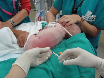
Lateral positioning of patient for spinal anesthesia
-
An alternative position for infants is to have an assistant hold the patient in a sitting position with the hips flexed and the head flexed forward. Cooperative adolescent patients can assume this sitting position themselves with a trained assistant facing them for support. The advantage of the sitting position is to increase the CSF pressure in the lumbar region and improve CSF flow through the spinal needle.

3.3 Surface Anatomy
Important surface anatomy landmarks that should be identified prior to sonographic assessment of the spine include:
-
Spinous processes to ascertain midline and to assess for abnormal spine curvature: Due to delayed fusion in neonates and infants, these may be palpable as two adjacent bony landmarks.
-
Iliac crests: An imaginary line between the anterior iliac crests, commonly known as the intercristal (or Truffier’s) line, will cross the L5–S1 interspace in neonates and infants less than 1 year old and L4–L5 in older children (Fig. 34.2).
Fig. 34.2 
Surface anatomy for pediatric spinal anesthesia. White and black lines indicate positions of iliac crests and intercristal line for neonates and children over 1 year old, respectively
-
Shoulders: Ensure that the left shoulder is not rotated forward and that both shoulders remain square to the bed. This will help ensure effective upper trunk flexion and alignment of the thoracic and lumbar spine, which may help with eventual dural puncture success.

Optimal positioning cannot be overemphasized and, once established, the Tegaderm® dressing should be removed and the residual gel wiped off. Prior to the spinal attempt, a sonographic assessment of the lumbar spine should be completed.
3.4 Sonographic Assessment
-
Sonographic assessment for spinal anesthesia is similar to that for lumbar epidural anesthesia (see Fig. 33.11). High-frequency (10–13 Hz) probes produce excellent resolution in small children and infants; however, in adolescents, the depth of neuraxial structures may necessitate a lower-frequency probe such as a curvilinear 2–6 MHz probe to gain adequate signal penetration.
-
A wider footprint linear-array transducer (10–13 MHz) allows for an excellent median or paramedian longitudinal view. Occasionally, a paramedian longitudinal view provides better detail, but this usually adds little to a median view in neonates and infants that are less than 6 months of age.
-
Expect the sonoanatomy to be excellent (>80 %) in children under 3 months of age, but it will gradually decline in quality to approximately 30–40 % by 9 months of age.
-
There are significant benefits to completing a transverse and median or paramedian view of the lumbar spine prior to performing the spinal anesthetic. This allows one to:
-
Identify the lumbar and sacral levels prior to dural puncture.
-
Delineate the spinous processes and ideal needle trajectory.
-
Identify the conus medullaris to be confident that dural puncture is below the termination of the cord.
-
Estimate the depth to the subarachnoid space; the distance between skin and the dura can be estimated, which in neonates and infants can be narrow (6–8 mm) [6]. If significant pressure is used with the probe during the sonographic assessment, the estimated depth may be erroneous.
-
-
Recently, it was demonstrated that real-time color flow Doppler ultrasound can be used to distinguish epidural injection from intrathecal injection (i.e., epidural injection produces a positive signal; intrathecal produces no signal) (see Fig. 33.22) [14].
Clinical Pearls
-
Ultrasound may be useful for pre-scanning.
-
Spinal anesthesia may be completed under real-time ultrasound guidance, but the merit of this approach is uncertain and may increase the risk of contamination due to the required extra equipment and personnel.
3.5 Nerve Stimulation Technique
The use of nerve stimulation to assist for spinal anesthesia in pediatric patients is not a common practice and therefore will not be described. However, the electrical epidural stimulation test can assist in distinguishing the epidural space (>1 mA) from the intrathecal space (<1 mA) when using an insulated needle (see Chap. 2) [15].
4 Equipment and Spinal Needle
-
Similar to all regional anesthetic blocks, there should be strict adherence to aseptic technique.
-
An absorbent pad should be placed between the warming blanket and the patient prior to the spinal anesthetic. Following the successful injection of the spinal anesthetic, the patient will occasionally have a bowel movement; an absorbent pad may be used to soak up these liquids that may otherwise inadvertently cool the patient over the course of the procedure.
-
Once in position for the spinal, the skin should be prepped with a 2 % chlorhexidine gluconate and 70 % isopropyl alcohol solution and allowed to dry.
-
A sterile clear plastic drape should be used, and a spinal needle and syringe containing the spinal anesthetic should be readily available.
-
Ensure comfortable ergonomics for both the anesthesiologist and the assistant holding the patient. We suggest that the anesthesiologist sit for stability and improved dexterity when attempting a spinal on an awake younger pediatric patient in lateral decubitus position.
-
If no topical gel or cream is used, infiltrate the skin with lidocaine 1 % using a 27G–30G needle prior to using the spinal needle.
4.1 Needles
-
The lumbar puncture is performed using a midline approach, preferably with a short 25G–27G styletted spinal needle. The type of needle has not been shown to have an effect on success or postspinal complications in the pediatric population [16, 17]. However, a smaller needle size could reduce the risk of post-dural puncture headache which is difficult to assess in this population.
-
Various types of spinal needles are available in pediatric sizes. Our approach is to use a 2.5 cm, 25G pencil-point needle (Pencan® Paed, B.Braun, Melsungen, Germany) and, if not successful, a 3.8 cm, 22G Quincke spinal needle. An introducer is not necessary in neonates and young infants.
-
In children, the ligamentum flavum is soft, and a distinctive “pop” may not be appreciated when the dura is punctured.
-
It is important to remove the stylet intermittently and examine for CSF flow. Initial CSF may be slightly blood tinged; ensure continued flow of clear fluid prior to injection of the anesthetic.
-
In neonates and young infants, use a 1 mL syringe (tuberculin syringe with clear gradations) to inject the drug slowly. A good rule of thumb is to inject over a 15–20 s period while avoiding the barbotage method as it may result in unacceptably high levels of motor blockade.
-
Once the subarachnoid block is performed, avoid elevating the legs or lower trunk. This will help to prevent cephalad spread of local anesthetic and is especially important during the application of the return pad which is typically fixed to the backs of neonates and young infants.
5 Local Anesthetics
-
Many drugs have been used for pediatric spinal anesthesia in variable doses for various surgical procedures. These drugs have been used as sole agents and also in combination with sedation and general anesthesia. Intrathecal agents used in the pediatric population include bupivacaine, tetracaine, lidocaine, ropivacaine, and levobupivacaine; adjuvants include morphine, fentanyl, clonidine, epinephrine, neostigmine, and dextrose.
-
The commonly used local anesthetics for pediatric spinal anesthesia include bupivacaine and tetracaine. Generally, a dose of 0.4–1 mg/kg of tetracaine or bupivacaine for spinal anesthesia will offer favorable surgical anesthesia. Higher doses per kg are preferred in the pediatric population, but the risk of a total spinal is rare as long as the procedure is carried out diligently. At our institution, the drug of choice is preservative-free plain bupivacaine 0.5 %. In neonates and infants weighing 5 kg or less, preservative-free plain bupivacaine 0.5 %, 1 mg/kg (0.2 mL/kg) is an effective dose that will provide 60 min of surgical anesthesia for inguinal hernia repair. Unfortunately, data for children outside the neonatal and infant stages are limited. As a general guide, the following suggested doses may be used:
-
0.3–0.5 mg/kg bupivacaine 0.5 % for children 2 months to 12 years of age
-
0.3–0.4 mg/kg hyperbaric tetracaine in children aged 12 weeks to 2 years
-
0.2–0.3 mg/kg hyperbaric tetracaine in older children of >2 years
-
5.1 Adjuvants
-
Clonidine (1 μg/kg) added to bupivacaine (1 mg/kg) has been used in spinal anesthesia in neonates and infants weighing 5 kg or less and provides almost twice the duration of spinal anesthesia when compared to local anesthetic alone [18]. Raising the intrathecal clonidine dose to 2 μg/kg provided no added benefit with propensity for transient drops in blood pressures intraoperatively and increased sedation in the postoperative period in this age group.
-
The use of intravenous caffeine (5–10 mg/kg) has been shown to prevent potential apnea in the postoperative period, especially if clonidine is used in the spinal anesthetic solution [18, 19].
-
An epinephrine washout of a tuberculin syringe may be preferred to a standard dose of intrathecal epinephrine (e.g., 0.01 mL/kg of 1:100,000 diluted epinephrine) for extending spinal block duration.
-
When compared to a eubaric solution, hyperbaric solution with dextrose does not seem to alter the duration of the spinal block in children.
See Table 34.1 for a summary of suggested local anesthetics and adjuvants.
6 Assessment of the Block Level
-
Assessment of the sensory and motor block can be challenging, especially in neonates, small children, and sedated patients.
-
In infants, response to cold stimuli (e.g., ice wrapped in a glove or an alcohol swab) can be used.
-
A Bromage score (see Table 34.2) [20], which is the gold standard, can usually be obtained for children greater than 2 years of age.
Table 34.2 Bromage scale for spinal block assessment -
If a rapidly rising level of blockade is noted, the patient may be placed in reverse Trendelenburg position to prevent further cephalad spread of local anesthetic.
7 Complications
-
The most common complications include multiple attempts, sensory and motor block failure requiring supplemental anesthetic, and surgical procedure outlasting the block.
-
Other less common complications include bleeding and hematoma, infection, allergic reaction, local anesthetic toxicity, cardiovascular complications, and nerve injury. The risk of methemoglobinemia is present with the use of tetracaine.
-
Although possible complications include post-dural puncture headache and transient radicular symptoms, these are less commonly reported in children.
8 Current Literature in Ultrasound-Guided Approaches
There is limited literature regarding the use of ultrasound for spinal anesthesia in the pediatric population, as the landmark technique has traditionally been used with success in children. However, as discussed above, ultrasound has potential value in identifying neuraxial structures and verifying the presence of anatomic abnormalities if they are present. Koo et al. [21] demonstrated that ultrasound could be used on children with urogenital abnormalities to identify occult spinal dysraphism. Further discussion of the use of pre-procedural ultrasound scanning for neuraxial blocks is found in a review by Chin and Perlas [22].
9 Case Study
Case Study: Spinal Anesthetic (Contributed by A. Spencer)
A 56-day-old baby presented for repair of bilateral inguinal hernias. This triplet was born at 30 weeks gestational age and, at time of surgery, weighed 2.48 kg. After birth, the patient remained in the neonatal intensive care unit for 5 weeks due to apneic episodes associated with bradycardia and due to dietary issues and was ultimately discharged home at 36 weeks corrected age. She had no known drug allergies and was on no regular medications except for daily iron and multivitamins.
Thirty minutes prior to the spinal anesthetic, Ametop gel (tetracaine 4 %) was applied to the L3–L5 lumbar spinal level and covered with a Tegaderm® dressing. After induction with sevoflurane (up to 4 %), the patient was placed in left lateral decubitus position and held in fetal position by an operative nurse. The dressing was removed and the residual gel wiped off, and the skin was prepped with a 2 % chlorhexidine gluconate and 70 % isopropyl alcohol swab. A linear-array transducer (10–13 MHz) was used to image the patient’s spine in a paramedian longitudinal view (Fig. 34.3). An assessment of the spinous processes, identification of lumbar levels, and assessment of the depth to the posterior dura and spinal cord and tip of the conus medullaris were completed. The sonogram of the lumbosacral region was then used to mark an entry level that would be below the conus. A 2.5 cm, 25G styletted spinal needle was used for dural puncture. Once cerebrospinal solution was found, 0.2 mL/kg (for a total volume of 0.5 mL) of preservative-free bupivacaine 0.5 % was slowly injected over 20 s. Block duration was 70 min; duration of surgery was 50 min.

Paramedian longitudinal ultrasound scan of neuraxial structures in an infant (see “Case Study” for details)
Acetaminophen 15 mg/kg p.o. liquid was given preoperatively and q6h postoperatively for 48 h. The patient was comfortable 30 min post-op. The patient spent the next 24 h under observation in the pediatric intensive care unit due to her age and prematurity and history of apneas and bradycardia. This observation period was uneventful, and the patient was discharged home the next day.
References
Frawley G, Ingelmo P. Spinal anaesthesia in the neonate. Best Pract Res Clin Anaesthesiol. 2010;24(3):337–51.
Bainbridge WS. A report of twelve operations on infants and young children using spinal analgesia. Arch Pediatr. 1901;18:570–4.
Gray HT. A study of spinal anaesthesia in children and infants. Lancet. 1909;2:913–7.
Abajian JC, Mellish RW, Browne AF, Perkins FM, Lambert DH, Mazuzan Jr JE. Spinal anesthesia for surgery in the high-risk infant. Anesth Analg. 1984;63:359–62.
Kesler H, Dias M, Kalapos P. Termination of the normal conus medullaris in children: a whole-spine magnetic resonance imaging study. Neruosurg Focus. 2007;23:E7.
Arthurs MM, Zubier M, Tooley J, Kelsall W. Ultrasonographic determination of neonatal spinal canal depth. Arch Dis Child Fetal Neonatal Ed. 2008;93:F451–4.
Bonadio W, Smith D, Metrou M, et al. Estimating lumbar-puncture depth in children. N Engl J Med. 1988;319:952–3.
Shenkman Z, Rathaus V, Jedeikin R, et al. The distance from the skin to the subarachnoid space can be predicted in premature and former-premature infants. Can J Anaesth. 2004;51:160–2.
Cote CJ, Lerman J, Todres ID. A practice of anesthesia for infants and children. Saunders, Elsevier: Philadelphia; 2009. Chapter 42 regional anesthesia, p. 877.
Cote CJ, Zaslavsky A, Downes JJ, Kurth CD, Welborn LG, Warner LO, Malviya SV. Postoperative apnea in former preterm infants after inguinal herniorrhaphy. A combined analysis. Anesthesiology. 1995;82:809–22.
Tobias JD. Spinal anaesthesia in infants and children. Pediatr Anesth. 2000;10:5–16.
Andrew M, Paes B, Milner R, Johnston M, Mitchell L, Tollefsen DM, Castle V, Powers P. Development of the human coagulation system in the healthy premature infant. Blood. 1988;72:1651–7.
De Saint Blanquat L, Simon L, Laplace C, Egu JF, Hamza J. Preoperative coagulation tests in former preterm infants undergoing spinal anaesthesia. Pediatr Anesth. 2002;12(4):304–7.
Tsui B, Leipoldt C, Desai S. Color flow Doppler ultrasonography can distinguish caudal epidural injection from intrathecal injection. Anesth Analg. 2013;116(6):1376–9.
Tsui BC, Wagner AM, Cunningham K, Perry S, Desai S, Seal R. Can continuous low current electrical stimulation distinguish insulated needle position in the epidural and intrathecal spaces in pediatric patients? Pediatr Anesth. 2005;15(11):959–63.
Kokki H, Heikkinen M, Turunen M, et al. Needle design does not affect the success rate of spinal anaesthesia or the incidence of postpuncture complications in children. Acta Anaesthesiol Scand. 2000;44:210–3.
Kokki H, Salonvaara M, Herrgard E, Onen P. Postdural puncture headache is not an age-related symptom in children: a prospective, open-randomized, parallel group study comparing a 22-gauge Quincke with a 22-gauge Whitacre needle. Pediatr Anesth. 1999;9:429–34.
Rochette A, Raux O, Troncin R, Dadure C, Verdier R, Capdevilla X. Clonidine prolongs spinal anesthesia in newborns: a prospective dose-ranging study. Anesth Analg. 2004;98(1):56–9.
Henderson-Smart DJ, Steer PA. Prophylactic caffeine to prevent postoperative apnoea following general anaesthesia in preterm infants. Cochrane Database Syst Rev. 2001;4:CD000048.
Bromage PR. A comparison of the hydrochloride and carbon dioxide salts of lidocaine and prilocaine in epidural analgesia. Acta Anaesthesiol Scand Suppl. 1965;16:55–69.
Koo BN, Hong JY, Song HT, Kim JM, Kil HK. Ultrasonography reveals a high prevalence of lower spinal dysraphism in children with urogenital anomalies. Acta Anaesthesiol Scand. 2012;56:624–8.
Chin KJ, Perlas A. Ultrasonography of the lumbar spine for neuraxial and lumbar plexus blocks. Curr Opin Anaesthesiol. 2011;24:567–72.
Suggested Reading
Dalens BJ. Spinal anesthesia. In: Dalens BJ, editor. Pediatric regional anesthesia. Boca Raton: CRC Press; 1990. p. 417–35.
Ganesh A, Kim A, Casale P, Cucchiaro G. Low-dose intrathecal morphine for postoperative analgesia in children. Anesth Analg. 2007;104:271–6.
López T, Sánchez FJ, Garzón JC, Muriel C. Spinal anesthesia in pediatric patients. Minerva Anestesiol. 2012;78:78–87.
Suresh S, Polaner DM, Cote CJ. Regional Anesthesia. In: Cote CJ, Lerman J, Anderson BJ, Eds. 5th ed. Philadelphia: WB Saunders; 2013. p. 835–79.
Puncuh F, Lampugnani E, Kokki H. Use of spinal anaesthesia in paediatric patients: a single centre experience with 1132 cases. Pediatr Anesth. 2004;14:564–7.
Rowney DA, Doyle E. Epidural and subarachnoid blockade in children. Anaesthesia. 1998;53:980–1001.
Saint-Maurice C. Spinal anesthesia. In: Dalens B, editor. Regional anesthesia in infants, children, and adolescents. Philadelphia: Lippincott Williams & Wilkins; 1995. p. 261–73.
Troncin R, Dadure C. Paediatric spinal anaesthesia. Update Anaesth. 2009;25:20–4.
Author information
Authors and Affiliations
Corresponding author
Editor information
Editors and Affiliations
Rights and permissions
Copyright information
© 2016 Springer Science+Business Media New York
About this chapter
Cite this chapter
Spencer, A.O., Suresh, S., Tsui, B.C.H. (2016). Spinal Anesthesia. In: Tsui, B., Suresh, S. (eds) Pediatric Atlas of Ultrasound- and Nerve Stimulation-Guided Regional Anesthesia. Springer, New York, NY. https://doi.org/10.1007/978-0-387-79964-3_34
Download citation
DOI: https://doi.org/10.1007/978-0-387-79964-3_34
Publisher Name: Springer, New York, NY
Print ISBN: 978-0-387-79963-6
Online ISBN: 978-0-387-79964-3
eBook Packages: MedicineMedicine (R0)