
Abstract
Regional anesthesia of the head and neck depends primarily on local infiltration and/or specific nerve blocks of terminal sensory branches (primarily of the fifth cranial (trigeminal) nerve) or of the cervical nerve roots C2–C4. Placement of these blocks depends on identification of reliable anatomical landmarks. Elicitation of paresthesia is generally not practical for nerve localization, since most pediatric patients are under sedation or general anesthesia while the block is performed. With the exception of the ultrasound-guided suprazygomatic maxillary nerve block, techniques using nerve stimulation or ultrasound imaging are not routinely used for facial blocks. Therefore, the description of techniques in this chapter will deviate from other chapters in which there is greater reliance on nerve localization modalities using nerve stimulation and ultrasound imaging. This section will focus on regional techniques commonly employed by anesthesiologists for perioperative and postoperative pain control in neurosurgery, plastic, and otolaryngology surgeries. Regional anesthesia techniques described in this chapter include superficial (transcutaneous) blocks to the main branches of the trigeminal nerve; intraoral blockade of the infraorbital, mental, and greater palatine nerves; as well as deeper nerve blocks of the maxillary and mandibular nerves. Uncommon regional blocks in pediatric patients, including ophthalmic, intraoral dental, and airway blocks, are not described.
Access provided by Autonomous University of Puebla. Download chapter PDF
Keywords
- Trigeminal nerve
- Supraorbital nerve block
- Infraorbital nerve block
- Mental nerve block
- Greater palatine nerve block
1 Indications
Blockade of the trigeminal nerve – the fifth cranial nerve – targets its three major branches (Fig. 9.1), the ophthalmic (V1, sensory), the maxillary (V2, sensory), and the mandibular (V3, sensory and motor to the muscles of mastication), and provides anesthesia to the anterior portion of the scalp, face, and much of the oral cavity. Some common clinical uses of trigeminal nerve block include (for a more comprehensive list, see Table 15.1):
-
Ophthalmic nerve block (V1: supraorbital, supratrochlear):
-
Frontal craniotomies
-
Excision of scalp nevus
-
-
Maxillary nerve block (V2: infraorbital, greater palatine):
-
Cleft palate surgery (maxillary, greater palatine, lesser palatine, nasopalatine)
-
Cleft lip surgery (infraorbital)
-
Nasal septum repair (infraorbital)
-
Endoscopic sinus surgery (infraorbital)
-
Surgical repair of soft tissue injury of the face (infraorbital)
-
Mandibular nerve block (V3: mental)
-
Lower lip repair (mental)
-
2 Block Techniques
The most comprehensive blockade of the trigeminal nerve targets the central ganglion. This block is usually performed by neurosurgeons under fluoroscopic guidance to treat disabling trigeminal neuralgia. Few anesthesiologists perform this technically difficult block, and it will not be described in detail here.
2.1 Superficial Transcutaneous Approach to Trigeminal Nerve Blocks: Supraorbital, Infraorbital, and Mental Nerve Block
The trigeminal block can be easily performed by injection of the three individual terminal superficial branches through a landmark-based approach using palpation of their respective foramina (Fig. 15.1). These bony landmarks are usually sufficient themselves for routine anesthetic purposes although ultrasound imaging may prove useful for locating them in some cases. An additional block of the supratrochlear nerve (terminal nerve of ophthalmic branch) is required if the field of anesthesia is to cross the midline of the forehead.

Patient positioning and surface landmarks for superficial trigeminal nerve blocks in a 4-year-old child
2.1.1 Patient Positioning
-
The patient is positioned lying supine.
-
The patient’s head may be placed to rest on a donut.
2.1.2 Landmarks and Surface Anatomy
-
Each nerve is closely associated with a readily palpable foramen. Figure 15.2 illustrates the cross-sectional anatomy of each respective foramen, captured by Visible Human Visualization Software (VHVS) and MRI.
Fig. 15.2 
VHVS and MRI of (a) supraorbital, (b) infraorbital, and (c) mental foramina
-
The nerves are too superficial to visualize well with ultrasound. However, ultrasound can be used to identify the foramina by scanning sagittally in the medial to lateral direction [1]. The foramina create discontinuity in the hyperechoic line of the bone. The absence of hyperechogenic bony structure indicates the position of the foramen (Fig. 15.3). Using this method, the foramina can be landmarked and marked on the skin to facilitate easier identification of the needle insertion site. Color Doppler can also be used to verify the locations of the foramina by imaging the blood vessels associated with each foramen; however, these vessels are small and are often difficult to visualize.
Fig. 15.3 
Ultrasound images with color Doppler demonstrating discontinuity in the hyperechogenic line at the supraorbital (a), infraorbital (b), and mental (c) foramina. Ultrasound probe positioning is indicated by blue bars in the center panel: “1” represents starting position and “2” represents final position over the foramen
-
All branches of the trigeminal nerve have been reported to lie in the same sagittal plane on each side of the face for adolescent and adult patients at a distance of approximately 2.5 cm lateral to the midsagittal line passing through the pupil [2]. Some reports indicate that accessory or double foramina may exist [3, 4], but unless one is attempting to enter the foramen while performing the nerve block, this should not affect clinical practice.
-
The supraorbital nerve enters the facial skeleton through the supraorbital foramen, which is located in the midsagittal plane at the level of the pupil. The supratrochlear nerve is located medial to the supraorbital foramen and can be found closer to the midfacial sagittal plane (Fig. 15.3a). The supraorbital notch is easily palpated at the medial upper angle of the orbit. Palpate the roof of the orbital rim starting from the midline. The more medial supratrochlear nerve is located at the upper internal angle of the orbital rim. Eipe et al. [5] describe their point of needle insertion as the intersection of a vertical line through the pupil of the eye and a horizontal line through the ala of the nose in their study of twenty children above 12 years of age.
-
The infraorbital notch (Fig. 15.3b) can be palpated easily along the floor of the orbital rim in children but can be difficult in the neonate due to the developing craniofacial skeletal configurations. If the foramen that exists inferior to the orbital rim cannot be palpated directly, it can be sought by gently probing with a small-gauge needle or found using surface landmarks as a guide. Alternatively, a simple mathematical formula can be utilized (distance from the midline = 21 mm + 0.5 × age (in years)) [2].
-
The mental nerve emerges from the mental foramen (Fig. 15.3c), which lies inferior to the outer lip at the level of the first premolar, midway between the upper and lower borders of the mandible [6].
-


2.1.3 Considerations and Needle Insertion Technique (Infraorbital Nerve Block)
-
Generally, short, 25G–30G hypodermic needles will be suitable for these blocks, provided that one is not trying to enter the foramen directly since this may require a larger needle for accurate placement. Longer needles are essential for:
-
Intraoral approaches to the infraorbital nerve in older children and adolescents (the infraorbital foramen may be located 2–3 cm away from the gingival sulcus)
-
Suprazygomatic maxillary nerve blocks in children of any age (depth of the pterygopalatine fossa from the superior junction of the zygoma and posterior orbital rim has been reported to be approximately 4 cm even in infants and young children)
-
Any approach to the pterygopalatine fossa from intra- or extraoral routes in children of all ages
-
-
Most exiting foramina communicate directly with the inferior orbital fissure. Thus, when using techniques that require injection of local anesthetic into the foramen itself, orbital contents may be injured inadvertently if one is not conscious of the depths and anatomic characteristics of the various canals where the nerves exit the facial skeleton.
-
Intraforaminal approaches are popular in many countries, but they have been reported to result in a higher incidence of paresthesia. Careful attention to injection pressure is critically important if the needle has entered the infraorbital canal itself. When placing the needle in these small foramina, the injection pressure must not be excessive, thereby minimizing trauma to the nerve. Intraforaminal approaches are not recommended for beginners since the basic approaches, when performed correctly, will provide excellent anesthesia and eliminate any risk of major complications.
-
When determining the optimal technique for infraorbital nerve block, the choice of an extraoral percutaneous route or an intraoral route (see below) must be made. While many practitioners advocate one technique over the other, either approach is appropriate, provided that the practitioner understands the benefits and limitations of the various approaches:
-
The extraoral percutaneous approach is relatively simple, and a supervisor and surgical team can visualize the injection and confirm where both the needle and local anesthesia are being placed.
-
The intraoral approach has been reported to be less painful for the patient [7]. In our opinion, the ability to supervise accurate needle placement and exact area of local anesthetic placement may be limited to the individual performing the block, particularly in small infants with cleft lip and palate defects.
-
-
Before the needle is introduced, the location and anatomic characteristics of the infraorbital foramen and the course of the exiting nerve branches should be studied. Understanding the shape and course of the foramen will help the practitioner decide how best to enter or, in most cases, avoid entering the foramen.
-
The infraorbital nerve travels from the foramen rotundum through the infraorbital canal and exits the facial skeleton through the infraorbital foramen. When examining the skull in the anterior plane, the infraorbital canal runs in a nasal-to-temporal direction; this directional information will help to plan entry or avoidance of the foramen.
-
If one plans on entering the foramen, then a nasal-temporal needle direction is recommended. For novice practitioners, a temporal-nasal needle direction will make it impossible to enter the canal. Any sagittally directed needle risks entering the foramen and should be avoided.
-
In infants, the infraorbital foramen will lie at the level of the nasal ala. The needle insertion point and location of the infraorbital foramen have been reported to be at the midpoint of a line drawn from the lateral orbital rim to the lateral corner of the mouth [8]. Due to the development of the craniofacial skeleton, a better surface landmark in older children is the intersection of a line drawn from the lateral orbital canthus to the nasal ala and another line passing sagittally through the medial edge (limbus) of the iris [9]. The foramen represents the initial point at which the infraorbital nerve starts to branch into its peripheral divisions.
-
-
The branches of the infraorbital nerve include the inferior palpebral, external nasal, internal nasal, and superior labial branch (which has a medial and lateral division). Only 40 % of the time do the nerves exit as separate branches [10]; the other 60 % of the time, the nerves exit as a network. Regardless, it is critical to remember that all the nerves travel in a lateral-to-medial direction toward the nasal ala. Successful nerve blockade is achieved when the local anesthesia spread matches these branching patterns. By understanding this principle, one can use visualization to determine when an adequate dose is achieved.
2.1.4 Needle Insertion Techniques: Infraorbital Nerve and Branches
-
Mark the skin with landmarks: the easiest landmark to identify in almost all ages is a point at the intersection of a line passing through the orbit at the vertical level of the limbic sagittal line and a line drawn from the lateral edge of the orbit to the nasal ala rim. At this point, the infraorbital foramen may be palpated.
-
Mark the foramen’s location, and place the index finger of the hand contralateral to the side to be blocked on the superior aspect of the eyelid of the side to be blocked (e.g., left index finger on the child’s right eyelid). This will enable the practitioner to hold the syringe like a pencil and perform accurate needle placement. Injection with the non-dominant hand requires practice but is worth the effort to improve accuracy.
-
With the free hand’s index finger, the patient’s eye can be examined for abnormalities and to confirm that stage 2 anesthesia is not present. Following this, place the index finger below the orbit in the area of the infraorbital notch. Take care not to push the eye as this may cause a bradycardiac response.
-
Lowering the eyelid and placing the finger on the inferior orbital rim can prevent local anesthetic from spreading in a superior direction, which is not necessary unless blockade of the inferior palpebral branch is needed for surgery. This will also help to prevent spread of local anesthetic to the loose soft tissues of the eye, which may lead to bruising and a black eye.
-
The needle is inserted in a temporal-to-nasal direction, directly superior and lateral to the marked infraorbital foramen. Entering a small distance away from this mark will help ensure needle contact with the bone in the area of the foramen itself. The goal is to not enter the infraorbital foramen directly but to place the needle in proximity to the infraorbital foramen when contact with the maxilla occurs.
-
Many authors describe backing the needle off the bone at this point. This is unnecessary but can be done if desired to decrease the risk of nerve injury. After aspiration of the syringe, inject enough local anesthetic to ensure spread to the nasal ala rim, thereby covering all nerve branching patterns. If local anesthetic tracks in any other direction, the block may not be accurate, and the needle should be repositioned.
Advanced Approaches
-
Advanced extraoral approaches include modifying the above technique and using a needle insertion point either at the infraorbital rim in the area of the infraorbital notch itself or entry of the infraorbital foramen directly. These superiorly based techniques are best performed in older children or adolescents undergoing awake surgery and allow the block to be performed relatively rapidly using small volumes of local anesthetic. This approach can be performed with a smaller volume of a more concentrated local anesthetic solution and may work at volumes of 0.25–1 mL per side to achieve adequate anesthesia. By moving the needle to the area immediately below the foramen, the needle tip can be placed directly under the fascia surrounding the infraorbital nerve as it exits the foramen while using the maxilla as a backstop. When done accurately, local anesthetic will be injected at the under-surface of the foramen, providing consistent and reliable anesthesia. An added benefit is that a gentle and quick injection can gain a scared awake child’s confidence.
-
Entering the infraorbital foramen is another advanced technique and is best performed if one remembers to change the needle direction to nasal-temporal. The foramen is located 7–8 mm below the orbital rim in adults and older children. When entering from a percutaneous nasal ala approach, a longer needle may be required. Zide [11] suggests to place the needle in the center of an imaginary triangle formed by the nasal labial fold, the nasal ala fold, and the foramen. Using this technique, block success was 100 % in adults when 1 mL local anesthetic was placed directly in the foramen, and no cases of nerve injury were found. To date, no studies have been done in pediatric patients using this technique.
2.1.5 V1: Ophthalmic Branches
-
The supraorbital and supratrochlear nerves are the terminal branches of V1 and can be blocked using one needle insertion with redirection (Fig. 15.4). After blocking the supraorbital nerve, the needle is withdrawn and redirected medially toward the supratrochlear foramen, where the supratrochlear nerve can be blocked.
Fig. 15.4 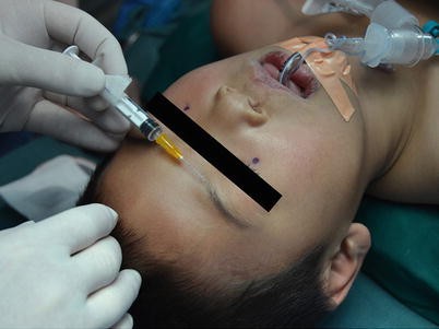
Needle insertion for transcutaneous supraorbital nerve block
-
The external nasal branch of the anterior ethmoid nerve and infratrochlear nerve innervates portions of the nasal bridge and the skin of the nasal ala, apex, and vestibule of the nose. These terminal branches of V1 can be blocked by insertion of a needle at the junction of the cartilaginous and bony portion of the nose on both lateral edges.
-
This block is typically required for surgeries such as nasal fracture repair, rhinoplasty, and any cleft repair that involves the surgeon working on the tip of the nasal area.
-
Typically, 0.5–1 mL is deposited in a superficial area at this superior nasal area using a small-gauge needle. In the case of bilateral cleft lip repair, it is important to place additional local anesthesia at the base of the nasal septum to ensure coverage of the prolabial fold. Typically, this area is covered by infraorbital nerve block via a branch of the internal nasal branch. However, this branch may not exist secondary to the bilateral cleft palate. This block is extremely painful for the awake patient and should be performed following all other blocks.
-
-
The mental canal angles medially and inferiorly; therefore, if the mental foramen cannot be palpated, subcutaneous injection can be performed above and lateral to the anticipated location of the mental foramen. This block can be performed intraorally or extraorally; anecdotal evidence suggests better success with the former approach, but no studies have been performed to confirm this.

2.1.6 Local Anesthetic Application
-
The choice of local anesthetic will depend on the purpose of the block and the duration of anesthesia required (e.g., 0.5–1 % lidocaine, 0.5–1 % mepivacaine for shorter procedures, and 0.2–0.5 % ropivacaine or bupivacaine for longer procedures). Textbooks will often quote the following doses for surgical anesthesia: 0.5–1.5 mL for infants, 2–3 mL for children, and 3–5 mL for adolescents. However, these volumes may be excessive, and in our experience, 1 mL or less is sufficient to block any infraorbital nerve provided the local anesthetic is placed accurately and one is attentive to adequate spread during block performance. Due to the vascularity of the face, epinephrine should always be used as an adjuvant to the local anesthetic to prolong duration of action. For diagnostic or therapeutic purposes, smaller volumes (0.5–1 mL) are recommended.
-
After needle insertion, the appropriate local anesthetic dose (e.g., 1–2 mL of 0.5–1 % lidocaine, or 0.5–1 mL of 0.125–0.25 % bupivacaine, or 0.2 % ropivacaine with epinephrine (1:200,000)) is injected slowly after aspiration.
-
Injection directly into the canals should be performed carefully to reduce the risk of neural injury.
-
Ensure that the local anesthetic reaches all the relevant branches in order to achieve adequate anesthesia. While many practitioners commonly think that they are blocking the infraorbital nerve directly, it is important to remember that the nerve branches as soon as it exits the skull.
-
Inaccurate placement of local anesthesia necessitates injection of larger volumes, which can be painful for awake and nonsedated patients. Inaccurate injection may also lead to partial blockade of maxillary nerve branches, which may fail to provide (1) adequate anesthesia to awake patients or (2) analgesia in the recovery room upon emergence.
Clinical Pearls
-
The blocks should be followed by local compression to prevent hematoma formation.
-
Intravascular injection is a rare but possible complication. Epinephrine (1:200,000) is a useful adjuvant that may help identify intravascular injection. Remember that the infraorbital artery lies in the middle of the nerve plexus, so aspiration of blood indicates that you are in the middle of the nerves. Repositioning the needle should lead to a successful block.
-
With infraorbital blocks, the child may bite their lip due to numbness. Thus, parents need to be warned of this effect and that feeding may be affected.
-
Skull nerve blocks can be used for craniotomy procedures and are also recommended to attenuate postoperative pain [12, 13]. The nerves blocked to achieve successful anesthesia for craniotomy include the supraorbital and supratrochlear nerves, the greater and lesser occipital nerves, the auriculotemporal nerves, and the greater auricular nerves. They can also be used for providing pain relief following minor surgical procedures on the forehead and scalp [14].
-
Supraorbital nerve blocks have been associated with a high requirement for supplementation, perhaps due to the anatomic variation of the nerve. The nerve may exit the skull undivided or its medial and lateral branches may exit separately.
2.2 Intraoral Approach to Trigeminal Nerve Blocks: Infraorbital, Mental, and Greater Palatine Nerves
Trigeminal nerves can also be blocked via the intraoral approach.
2.2.1 Patient Positioning
-
Supine with head resting on a donut.
-
Greater palatine nerve: to keep the mouth open during blockade of the greater palatine nerve, a bite block or, more appropriate, a Dingman mouth retractor is required.
2.2.2 Landmarks and Surface Anatomy
-
Infraorbital foramen: intraorally, locate the subsulcal groove at the level of the canine or first premolar (Fig. 15.5).
Fig. 15.5 
Surface anatomy and needle insertion for intraoral infraorbital nerve block
-
Mental foramen: the buccal mucosa of the first premolar is identified (Fig. 15.6).
Fig. 15.6 
Surface anatomy and needle direction for intraoral mental nerve block
-
Greater palatine foramen: in the intact palate, locate the greater palatine foramen medial and anterior to the first premolar in infants (Fig. 15.7) and second molar in adolescents. The nerve runs anterior to the foramen on the floor of the hard palate. In a patient with cleft palate, locate the greater palatine foramen by palpating the medial edge of the cleft palate. The end of the hard palate represents the level at which the greater palatine nerve exits the foramen and can be located consistently at this level. When palpating the medial bone edge, care must be taken to recognize the correct structures. Moving the finger randomly posterior risks palpating the hook of the hamulus rather than the true medial edge of the bone, which represents the level of the greater palatine foramen. Palpation of the hamulus will be too posterior, and injection here will result in ineffective anesthesia. To correct this, the finger should be moved toward the lateral edge of the palate, and a depression or groove will be felt, representing the foramen. This is the exit for the nerve and vascular pedicle that accompanies the nerve and supplies blood supply to the palatal tissues.
Fig. 15.7 
Surface anatomy and needle direction for greater palatine nerve block


2.2.3 Needle Insertion Technique
-
Intraoral infraorbital nerve: evert the upper lip and, using a ≥45° angle, insert a 23G–27G needle into the buccal mucosa in the subsulcal groove (Fig. 15.5). A longer needle is needed to reach the level of the foramen. External palpation at the foramen will help to prevent the needle from entering the globe of the eye.
-
Lateral needle placement or local anesthetic spread risks partial blockade. Placing local anesthetic too far laterally on the maxilla may result in blockade of the middle or anterior superior alveolar nerves; in this case, only dental – not surgical – anesthesia may be achieved.
-
Inaccurate needle direction may make location of the foramen difficult since the needle is traveling in a sagittal or nasal-to-temporal direction from the buccal sulcus.
-
-
Mental nerve: Evert the lower lip and insert a 25G–27G needle into the buccal mucosa between the canine and the first premolar (Fig. 15.6).
-
Greater palatine nerve: for patients with an intact palate, insert a 25G–27G needle into the mucosa anterior to the greater palatine foramen (Fig. 15.7). For cleft palate repairs, several separate blocks will be needed since the greater palatine nerve is only one of the nerves that innervate the cleft palate. The nasopalatine nerve, which is a terminal branch of the maxillary nerve, innervates the anterior or primary palate and the front four teeth and will need to be blocked separately. The soft palate, which is supplied by the lesser palatine nerve and a plexus of nerves that arise from the glossopharyngeal plexus in the oral cavity, also requires a separate injection.
-
When blocking these nerves, it is critical that the mouth be opened as widely as possible with the assistance of some type of mouth retractor such as a Dingman retractor. Having a wide open view allows both the practitioner and surgeon to have a full view of needle placement and prevent injury to the vascular pedicle.
-
After locating the greater palatine foramen, the injection is performed in a cross-mouth technique, which will allow everyone involved in the operation to view needle insertion and local anesthetic spread.
-
Use of epinephrine as an adjunct is critical so that the blanching of mucosa can help determine optimum local anesthetic spread.
-
Performing the injection at the beginning of surgery allows local anesthetic spread to be followed without any confusion from the surgical injection, which is typically performed for palate hemostasis and dissection of soft tissues. Ideally, the injection is performed in a gentle, single-shot technique without hunting for the foramen directly.
-
Ideally, the needle will contact the bone of the palate in a periforaminal location. Typically, 0.5–1 mL local anesthesia is injected. Spread in the hard palate only indicates successful injection; if the soft palate tissues begin to dissect, then the needle may be too far posterior and may need to be repositioned slightly anterior.
-
After the greater palatine nerve is surrounded by local anesthetic, the needle is inserted in a posterior direction about 3–5 mm posterior from the original injection position. The soft tissues that make up the soft palate pedicle are expanded by an additional 0.5–1 mL of local anesthetic. The lesser palatine foramen is too difficult to locate clinically since it is very lateral on the palate and extremely small (Fig. 15.8). Since a plexus of nerves innervates the soft palate, spreading the local anesthetic through the soft palate tissues will ensure posterior spread to reach the lesser palatine foramen. Medial spread will help anesthetize the glossopharyngeal components and help to ensure complete palate anesthesia.
Fig. 15.8 
Surface anatomy and needle direction for nasopalatine nerve block
-
-
The last nerve to block for palate anesthesia is the nasopalatine nerve (Fig. 15.8). It exits the nasopalatine foramen, which is located under the nasopalatine papilla in the front of the hard palate. The foramen is the entrance to a canal that travels in the direction of the tooth roots at a 45° angle.
-
A 25G–27G needle may be placed in this foramen; however, it is not uncommon to encounter difficulty entering the foramen, and some force may be required upon injection to ensure local anesthetic spread through the foramen.
-
If the cleft palate extends through the foramen, any injected local anesthesia will drip into the nasal vault and will not be effective. In this situation, it is possible to inject on both anterior surfaces of the palate to perform so-called partial palatal injections, which will anesthetize the anterior palate.
-
In some palate repairs, the vomer will also be used for the repair; additional local anesthesia should be placed here to ensure complete anesthesia of the surgical area.
-

2.2.4 Local Anesthetic Application
After aspiration, the appropriate local anesthetic dose (e.g., 1–2 mL of 0.5–1 % lidocaine, or 0.5–1 mL of 0.125–0.25 % bupivacaine, or 0.2 % ropivacaine with epinephrine 1:200,000) is slowly injected after aspiration.
2.3 Deep Trigeminal Nerve Blocks
Deep trigeminal nerve blocks are not commonly performed in the pediatric population; however, it is worth discussing the suprazygomatic maxillary nerve block, which has recently become popular for cleft palate repairs. These blocks have not been commonly performed, even in the adult population, most likely because of the deep insertion required to perform accurate nerve blockade and the potential for harm.
-
Deep trigeminal blocks are required when the superficial block of the infraorbital nerve does not produce adequate anesthesia or when complete maxillary or mandibular anesthesia is required.
-
Maxillary nerve block (often but not completely accurately referred to as sphenopalatine block) can be performed by a lateral approach to enter the sphenopalatine fissure. This can be achieved by approaching either above or below the zygomatic arch. Novice practitioners should exercise caution with this block since accurate needle placement requires insertion 3–4 cm deep into the skull base in a blind fashion. The recommended insertion point is at the junction of the superior zygoma and posterior orbit. The needle is inserted perpendicularly to contact the greater wing of the sphenoid at a depth of 2 cm. After bony contact, the needle is redirected 9° inferior and 20° anterior in the direction of the plane of the philtrum and is advanced 3–4 cm further to enter the pterygopalatine fossa (Fig. 15.9). A dose of 0.15 mL/kg is recommended after negative aspiration. The block is then repeated on the opposite side for complete maxillary nerve anesthesia.
Fig. 15.9 
Surface anatomy and needle direction for maxillary nerve block. (1) The needle is inserted perpendicularly to contact sphenoid; (2) the needle is redirected 9° inferior and then (3) 20° anterior; (4) the needle/syringe can hold its own position once the needle has entered the pterygopalatine fossa

The mandibular nerve can be blocked at the point where it leaves the cranium through the foramen ovale. The block can be done with an intraoral or extraoral approach through the intercondylar mandibular notch.
-
When using an extraoral approach, place the needle through the notch in a perpendicular fashion, and contact the pterygoid plate. Measure the depth and move the needle posterior to walk off the pterygoid plate. The needle should be advanced no further than the depth of the pterygoid plate. While no pediatric studies exist to recommend accurate dosing, 1–3 mL of local anesthetic should achieve adequate anesthesia since the needle is close to the foramen ovale and skull base.
Due to the depth of these blocks (see Figs. 15.10 and 15.11), they should be performed by practitioners with related and adequate experience.

VHVS and MRI images capturing the maxillary nerve during its course through the infraorbital groove and canal. This will be the location of a maxillary nerve block

VHVS and MRI of the mandibular nerve showing its position posterior to the maxillary nerve yet remaining medial to lateral pterygoid plate
3 Current Literature in Ultrasound-Guided Approaches
It has been reported that failure to achieve full anesthesia using traditional blocks of the trigeminal nerve is in the region of 22 % [15]. In pediatric patients, one reason for this may be that landmarks used to perform superficial trigeminal nerve blocks (particularly infraorbital) are absent or difficult to palpate in the neonate. Facial foramina can be localized accurately and reliably using ultrasound [1, 16], and this may provide an opportunity to improve success in these blocks. Tsui [1] described an ultrasound approach to locate the supraorbital, infraorbital, and mental foramina (see above). Identification of each foramen can be achieved using a high-resolution, short-footprint linear transducer; a disruption in the continuity of the bone will appear as scanning proceeds in a medial-to-lateral direction at the level of the foramina (Fig. 15.3). Color Doppler will prove useful to locate the respective artery within the foramen.
The effectiveness of trigeminal nerve block has been demonstrated in various studies. Ahuja et al. [17] showed a significant improvement in pain score in the infraorbital block group compared to normal saline infiltration in children scheduled for cleft lip repair. A double-blinded, randomized study also demonstrated a significant reduction in analgesia requirement and pain score in children who had an infraorbital nerve block compared to the conventional peri-incisional infiltration by the surgeon [18]. Recently, Mesnil et al. [19] observed the effectiveness of bilateral maxillary nerve blocks using a suprazygomatic approach, finding improved pain relief and a reduction in opioid consumption following cleft palate repair in infants. Furthermore, this approach minimizes the likelihood of entering the orbit and reduces the risk of vascular injury as the needle enters the infratemporal fossa and ultimately the sphenopalatine fossa in a superior-to-inferior direction. No significant difference was found in mandibular infiltration anesthesia and mandibular block for dental surgery in children [20]; however, in this study, mixed surgical cases with a relatively small sample size were investigated, which could introduce confounding factors.
The effectiveness of bilateral maxillary nerve blocks using a suprazygomatic approach with nerve stimulation [19] and ultrasound [21] has been reported. Using electrical stimulation, Mesnil et al. [19] showed that the disappearance of the temporal muscle twitch coincided with the needle’s tip in the pterygopalatine fossa where local anesthetic was injected. More recently, this group successfully published a randomized, double-blind study evaluating an ultrasound-guided suprazygomatic maxillary nerve block [21]. They used a linear array probe located in the infrazygomatic area to allow out-of-plane visualization of the needle tip and local anesthetic spread in the pterygopalatine fossa. Both these approaches allowed improved pain relief and a reduction in opioid consumption following cleft palate repair in infants.
4 Case Study
Greater Palatine Nerve Block (Provided by S. Suresh)
A 5-month-old female infant, 6.5 kg in weight, with a past medical history of cleft lip and cleft palate and mother with cleft lip, presented for cleft palate surgery. Hemoglobin was measured at 9.8 g%. No preadmission medications were given. A greater palatine nerve block was administered; briefly, the mouth gag was placed by the surgeon, after which the greater palatine foramen was identified in the hard palate (this usually corresponds to the second molar in an older patient with dentition), and 0.5 mL 0.25 % bupivacaine was injected with a 27G needle into the area anterior to the greater palatine foramen bilaterally (cf. Fig. 15.7). Duration of surgery was 2 h, 35 min; block duration was 8–10 h. No ultrasound imaging considerations were necessary, and no additional opioid was needed in recovery. Patient outcome was excellent and resulted in early discharge from PACU.
References
Tsui BC. Ultrasound imaging to localize foramina for superficial trigeminal nerve block. Can J Anesth. 2009;56:704–6.
Suresh S, Voronov P, Curran J. Infraorbital nerve block in children: a computerized tomographic measurement of the location of the infraorbital foramen. Reg Anesth Pain Med. 2006;31:211–4.
Canan S, Asim OM, Okan B, Ozek C, Alper M. Anatomic variations of the infraorbital foramen. Ann Plast Surg. 1999;43:613–7.
Thakur G, Thomas S, Thayil SC, Nair PP. Accessory mental foramen: a rare anatomical finding. BMJ Case Rep. 2011; 2011; bcr0920103326.
Eipe N, Choudhrie A, Pillai AD, Choudhrie R. Regional anesthesia for cleft lip repair: a preliminary study. Cleft Palate Craniofac J. 2006;43:138–41.
Voronov P, Suresh S. Head and neck blocks in children. Curr Opin Anaesthesiol. 2008;21:317–22.
Lynch MT, Syverud SA, Schwab RA, Jenkins JM, Edlich R. Comparison of intraoral and percutaneous approaches for infraorbital nerve block. Acad Emerg Med. 1994;1:514–9.
Bosenberg AT, Kimble FW. Infraorbital nerve block in neonates for cleft lip repair: anatomical study and clinical application. Br J Anaesth. 1995;74:506–8.
Wilhelmi BJ, Mowlavi A, Neumeister MW, Blackwell SJ. Facial fracture approaches with landmark ratios to predict the location of the infraorbital and supraorbital nerves: an anatomic study. J Craniofac Surg. 2003;14:473–7.
Hu KS, Kwak HH, Song WC, Kang HJ, Kim HC, Fontaine C, Kim HJ. Branching patterns of the infraorbital nerve and topography within the infraorbital space. J Craniofac Surg. 2006;17:1111–5.
Zide BM, Swift R. How to block and tackle the face. Plast Reconstr Surg. 1998;101:840–51.
Suresh S, Bellig G. Regional anesthesia in a very low-birth-weight neonate for a neurosurgical procedure. Reg Anesth Pain Med. 2004;29:58–9.
Uejima T, Suresh S. Ommaya and McComb reservoir placement in infants: can this be done with regional anesthesia? Pediatr Anesth. 2008;18:909–11.
Suresh S, Wagner AM. Scalp excisions: getting “ahead” of pain. Pediatr Dermatol. 2001;18:74–6.
Pascal J, Charier D, Perret D, Navez M, Auboyer C, Molliex S. Peripheral blocks of trigeminal nerve for facial soft-tissue surgery: learning from failures. Eur J Anaesthesiol. 2005;22:480–2.
Hannan L, Reader A, Nist R, Beck M, Meyers WJ. The use of ultrasound for guiding needle placement for inferior alveolar nerve blocks. Oral Surg Oral Med Oral Pathol Oral Radiol Endod. 1999;87:658–65.
Ahuja S, Datta A, Krishna A, Bhattacharya A. Infra-orbital nerve block for relief of postoperative pain following cleft lip surgery in infants. Anaesthesia. 1994;49:441–4.
Prabhu KP, Wig J, Grewal S. Bilateral infraorbital nerve block is superior to peri-incisional infiltration for analgesia after repair of cleft lip. Scand J Plast Reconstr Surg Hand Surg. 1999;33:83–7.
Mesnil M, Dadure C, Captier G, Raux O, Rochette A, Canaud N, Sauter M, Capdevila X. A new approach for peri-operative analgesia of cleft palate repair in infants: the bilateral suprazygomatic maxillary nerve block. Pediatr Anesth. 2010;20:343–9.
Yassen GH. Evaluation of mandibular infiltration versus mandibular block anaesthesia in treating primary canines in children. Int J Paediatr Dent. 2010;20:43–9.
Sola C, Raux O, Savath L, Macq C, Capdevila X, Dadure C. Ultrasound guidance characteristics and efficiency of suprazygomatic maxillary nerve blocks in infants: a descriptive prospective study. Pediatr Anesth. 2012;22:841–6.
Suggested Reading
Belvis D, Voronov P, Suresh S. Head and neck blocks in children. Tech Reg Anesth Pain Manag. 2007;11(4):208–14.
Dalens B. Blocks of the head, neck, and face. In: Dalens B, editor. Regional anesthesia in infants, children, and adolescents. Philadelphia: Lippincott Williams & Wilkins; 1995. p. 398–401.
Suresh S, Polaner DM, Cote CJ. Regional Anesthesia. In: Cote CJ, Lerman J, Anderson BJ, Eds. 5th ed. Philadelphia: WB Saunders; 2013. p. 835–79.
Suresh S, Voronov P. Head and neck blocks in infants, children, adolescents. Pediatr Anesth. 2012;22:81–7.
Author information
Authors and Affiliations
Corresponding author
Editor information
Editors and Affiliations
Rights and permissions
Copyright information
© 2016 Springer Science+Business Media New York
About this chapter
Cite this chapter
Merritt, G., Tsui, B.C.H. (2016). Trigeminal Nerve Blocks. In: Tsui, B., Suresh, S. (eds) Pediatric Atlas of Ultrasound- and Nerve Stimulation-Guided Regional Anesthesia. Springer, New York, NY. https://doi.org/10.1007/978-0-387-79964-3_15
Download citation
DOI: https://doi.org/10.1007/978-0-387-79964-3_15
Publisher Name: Springer, New York, NY
Print ISBN: 978-0-387-79963-6
Online ISBN: 978-0-387-79964-3
eBook Packages: MedicineMedicine (R0)